
In Vitro Differentiation of Human Liver Organoid Progenitors to Functional Liver Organoids
Section overview
- Human iPSC-derived Liver Organoids and Hepatic PDOs
- Materials
- Thawing liver progenitor organoids into 24-well plates
- Trypsinization-based passaging of liver progenitor organoids
- Differentiation of iPSC LPOs into mature liver organoids
- Differentiation of PDO LPOs into mature liver organoids
- Mature liver organoid characterization
- Related products
Human iPSC-derived Liver Organoids and Hepatic PDOs
As 3D cell culture models of the liver, hepatic organoids are used by researchers studying liver diseases, hepatic development and regeneration, metabolism, and more. Our organoid portfolio includes 3dGRO® Human iPSC-derived Liver Organoid Progenitors, which are derived from integration-free human iPSC and reprogrammed using Simplicon® RNA Reprogramming Technology. The human iPS cells were differentiated using a defined, multi-step differentiation protocol, yielding liver progenitor organoids (LPOs). The organoids are self-organizing 3D structures that express key markers such as HNF4a, SOX17, FOXA2 and SOX9, and display proper apical and basolateral polarization, as indicated by E-cadherin and ZO1 expression. Our portfolio also includes 3dGRO® Human Patient-Derived Liver Organoids (PDOs). The 3DGRO® normal liver organoid biobank consists of Immature Liver Organoids (ILO) which can be thawed, passaged and banked back. The ILO express markers for both hepatocytes and cholangiocytes, and when needed, they may be differentiated into Mature Liver Organoids (MLO), which are terminally differentiated.
Here we describe how to culture and differentiate our human iPSC-derived or patient-derived liver organoid progenitors to mature liver organoids containing both hepatocytes and cholangiocytes, as well as how to stain and characterize these organoids.
LPOs can be cultured and maintained long-term using 3dGRO® Human Liver Progenitor Organoid Expansion Medium (SCM313). To initiate maturation, LPOs are cultured for 7 days in Liver Organoid Differentiation Medium, followed by an additional 7-day culture in Hepatocyte Culture Medium (Lonza, CC-3198), resulting in functional MLOs containing hepatocytes and cholangiocytes.
Materials
- 3dGRO® Human Patient-Derived Liver Organoids (SCC570, SCC571)
- 3dGRO® Human iPSC-derived Liver Organoid Progenitors (SCC572)
- Corning Matrigel® Growth Factor Reduced (GFR) Basement Membrane Matrix (CLS356231)
- Basal Plus Medium (Composition: DMEM/F12 PLUS Basal Medium (SCM162) containing 5 mL penicillin-streptomycin (100X), 5 mL Ala-Gln (100X) and 5 mL HEPES (1 M)).
- 3dGRO® Human Liver Progenitor Organoid Expansion Medium (SCM313)
- Liver Organoid Differentiation Medium (see Table 1)
- HCM™ Hepatocyte Culture Medium Bulletkit® (Lonza, CC-3198)
- DMEM/F-12 PLUS Basal Media (SCM162)
- DMEM-High Glucose Medium (SLM-241)
- 1M HEPES Solution (H0887)
- Penicillin-Streptomycin (100X) (P0781)
- Ala-Gln, 200mM Solution (G8541)
- ROCK Inhibitor, Y-27632 (SCM075)
- Greiner CELLSTAR® 24-well tissue culture treated plates (M8812)
- 0.25% Trypsin-EDTA solution (T4049)
- Corning® Cell Recovery Solution (CLS354253)
- 1X D-PBS w/o Ca2+ and Mg2+ (D8537)
- Fetal Bovine Serum (ES009-M)
Please see data sheet for full comprehensive list of materials required for protocol.
Thawing liver progenitor organoids into 24-well plates
- Prior to thawing LPO, thaw sufficient GFR Matrigel® on ice for 12 domes at 50 μL per dome, plus a 5% overage (total: 630 μL).
Note: GFR Matrigel® polymerizes quickly at room temperature. Always keep it on ice post thaw. - Pre-warm a sterile, tissue culture-treated 24-well plate in a 37 °C incubator for 1-2 hours before use.
- Prepare Basal Plus Medium:
- In 500 mL of DMEM/F-12 PLUS Basal Medium (SCM162), add 5 mL penicillin-streptomycin (100X), 5 mL Ala-Gln (100X) and 5 mL of 1 M HEPES. Store at 2-8 °C for up to 6 months. The Basal Plus Medium is used for washing, passaging, and thawing LPO. Pre-warm the medium to room temperature for 15 minutes before use.
- Rapidly thaw the frozen vial of LPOs in a 37 °C bath. Immediately transfer contents to a 15 mL conical tube containing 9 mL of pre-warmed Basal Plus Medium. Handle Gently. Pipetting at this point should be as gentle as possible as organoids are very fragile at this stage.
- Rinse the cryovial with 1 mL Basal Plus Medium and add to the 15 mL conical tube.
- Centrifuge the 15 mL tube at 350 x g for 5 min at 25 °C.
- Carefully aspirate and discard the supernatant. Use a micropipette to remove any residual supernatant.
- Remove the pre-warmed 24-well tissue culture plate from the incubator.
- Gently resuspend the organoid pellet in 630 μL GFR Matrigel®. Avoid creating air bubbles.
- Dispense 50 μL Matrigel®/organoids suspension per well into 12-wells of the 24-Well plate. Let the Matrigel® domes set at room temperature for 2 min. Transfer the plate to a 37 °C, 5% CO2 incubator for 20 min to solidify.
- Carefully add 650 μL of 3dGRO® Human Liver Progenitor Organoid Expansion Medium (SCM313) to each well by dispensing along the wall to avoid disturbing the dome.
- Incubate at 37 °C, 5% CO2 in a humidified incubator overnight.
- Replace medium every 3-4 days or if the color changes to yellow.
Note: LPOs are typically ready for passage after 8-14 days. Post thaw recovery may be delayed if stored at −80 °C upon receipt. - Observe domes under the microscope and capture images on days 0, 2, 4, 8 and the day of passage. Figure 2 shows the density and morphology on Days 0 and 8. Figure 3 displays various LPO morphologies at Day 8.
- Passaging LPOs:
- Passage LPOs 10-14 days post thaw.
- After recovery, passage every 8-10 days using:
- Trypsinization Method: split 1:8-1:10
- Fragmentation Method: split 1:4

Figure 1.Diversity in ScLPO morphology. ScLPO can appear in different sizes, densities, and structures. Images are at 10X magnification.
Trypsinization-based passaging of liver progenitor organoids
Ensure that the organoids are healthy with minimal cell death and in the exponential growth phase before passaging using this method. There are several advantages to passaging liver progenitor organoids using the trypsinization method; each single cell or small cell cluster gives rise to one organoid, allowing the number of organoids to be estimated during seeding, and organoid size is more uniform, making them suitable for biobanking and for performing functional assays following differentiation.
- Prewarm the 24-well plates (for 1 hour), the Basal Plus Medium, DMEM with 10% FBS, 0.25% Trypsin EDTA (for 15 min) and 3dGRO® Human Liver Progenitor Organoid Expansion Medium (SCM313) at room temperature. Prior to passaging the LPOs, thaw enough GFR Matrigel® vials (1 hour before).
- Remove media from the wells. Wash Matrigel® domes with 500 μL DPBS (Ca2+ and Mg2+ free, room temperature). Aspirate the DPBS.
- Add 450 μL of 0.25% Trypsin EDTA to each well. Using a P1000 pipette, pipette up and down vigorously 5-6 times to release the organoids from the Matrigel® dome. This step facilitates the trypsinization of the organoids. Do not digest more than 12 wells from a 24-well plate per batch to avoid over-digestion. Excessive exposure to trypsin may reduce the viability of LPOs post passaging.
- Incubate the plates at 37 °C for 9 min. Remove plate from the 37 °C incubator and pipette each well up and down 5-6 times using a P1000 pipette to dissociate the organoids into single cells or small cell cluster. Add 1 mL of DMEM containing 10% FBS to each well to neutralize the trypsin. Transfer the contents of each well into a 15 mL tubes. If all 12 wells contain organoids, use two 15 mL tubes to accommodate the total volume.
- Centrifuge at 350 x g for 5 min at 4 °C. Aspirate the supernatant; add 10 mL of Basal Plus Medium to wash away the FBS.
- Centrifuge again at 350 x g for 5 min at 4 °C. Remove all the supernatant.
- Count cells or cell clusters using a cell counter. Calculate the number needed to seed 1500 cells or cell clusters per well.
- Resuspend gently the calculated number of cells or cell clusters in Matrigel®. Plate 50 μL Matrigel® dome into each well of a 24-well plate.
Example calculation: To seed 1500 cells or cell clusters per well across an entire 24-well plate, 36,000 cells are required. Resuspend the 36,000 cells in 1.2 mL of Matrigel® with an additional 5% overage. Mix thoroughly and dispense 50 μL of the mixture into each well. - Transfer the plate to a 37 °C, 5% CO2 incubator for 20 min to allow the Matrigel® to polymerize.
- Gently add 650 μL of 3dGRO® Human Liver Progenitor Organoid Expansion Medium (SCM313) to each well. To avoid disturbing the domes, dispense the medium against the side of each well.
- Replace the medium every 3–4 days, or sooner if the medium turns yellow.
- Monitor the domes under a microscope and capture images on Days 0, 2, 4, 6 and the day of passaging for documentation. Figure 4 shows the morphology of LPOs on various days following passaging via the trypsinization method.
- For biobanking, it is recommended to cryopreserve organoids on Days 6–7, when they are in the exponential growth phase, to ensure optimal recovery after thawing. For each biobank vial, pool organoids from approximately 2 wells (about 1000–1200 LPOs per well), resulting in a total of ~2000-2400 LPOs per vial.
Note: For more information on the cryopreservation protocol, see the data sheet.
- All growth factors must be prepared in DPBS-B (1X PBS with 0.1% BSA).
Note: To make 0.1% BSA solution, dilute sterile 10% BSA Stock Solution (A8806) in 1X PBS (TMS-012) and filter sterilize before use. - A83-01 (TGF-beta/Smad Inhibitor, Mol. Wt. 421.52 g/mol)
Dissolve 25 mg of A83-01 in 5.9 mL of sterile DMSO to prepare a 10 mM stock solution. Aliquot 10 μL portions and store at −20 °C for up to 1 year.
Important: Avoid repeated freeze-thaw cycles to preserve stability and activity. - Dexamethasone (Mol. Wt. 392.46 g/mol)
Dissolve 25 mg of Dexamethasone in 6.37 mL of sterile DMSO to make a 10 mM stock solution. Aliquot 15 μL portions and store at −20 °C for up to 1 year.
Important: Do not reuse thawed aliquots. Avoid repeated freeze-thaw cycles. - [Leu15]-Gastrin I Human (Mol. Wt. 2080.16 g/mol)
Dissolve 25 mg of [Leu15]-Gastrin I human in 2.4 mL of sterile 1X PBS containing 0.1% BSA to prepare a 100 μM stock solution. Aliquot 10 μL portions and store at −20 °C for up to 1 year.
Important: Do not reuse thawed aliquots. Avoid repeated freeze-thaw cycles.
LPO Differentiation Protocol
- Differentiation protocol
Liver progenitor organoids (LPOs) can be differentiated into mature liver organoids by culturing in Liver Organoid Differentiation Medium (see Table 1 for composition) for 7 days. This is followed by a 7-day maintenance phase in Hepatocyte Culture Medium (HCM™ Hepatocyte Culture Medium BulletKit®, Lonza, CC-3198). - Timing and seeding density
Initiate differentiation 10 days post-passaging of LPOs using the trypsinization method. Recommended seeding densities:- ~300 organoids/50 μL dome (24-well plate)
- ~25-45 organoids/10 μL dome (96-well plate)
- Differentiation notes
A cell death rate of approximately 5-10% is commonly observed during differentiation.
Important: Mature liver organoids do not expand or proliferate and therefore cannot be passaged. - Media changes
Change medium every 3-4 days, or sooner if the medium appears yellowish. - Post-differentiation maintenance
Mature liver organoids can be maintained in Hepatocyte Culture Medium (Lonza, CC-3198) for up to 1 month. During this time, they are suitable for downstream applications such as functional assays, DMPK studies and toxicology testing.
Differentiation of PDO LPOs into mature liver organoids
Preparation of liver PDO differentiation medium
Differentiation of immature liver organoids (ILO) into Mature Liver Organoids (MLO)
Important Notes:
- Mature liver organoids do not expand or proliferate and therefore cannot be passaged.
- ILO should be thawed and passaged at least twice before differentiation. Ideally, MLO should be freshly generated before any relevant assays and should be discarded following the experiment.
- Recommended seeding densities:
For 24 well plates ~150- 200 organoids/25 μL dome
For 96 well plates ~25-45 organoids/10 μL dome - A cell death rate of approximately 5-10% is commonly observed during differentiation.
- Media should be changed every 3-4 days, or sooner if the medium appears yellowish.
- Passage ILO following the protocol described before and seed Matrigel® matrix/ organoid domes.
- Culture domes in pre-differentiation medium (Liver PDO Expansion media + 25 ng/ ml BMP7, B1434-10UG) for 3-5 days.
- Following pre-differentiation, grow the organoids for 7 days in Liver PDO Differentiation Medium (see Table 1), as described above.
- Next, culture the organoids for 7 days in Hepatocyte Culture Medium (HCM™ Hepatocyte Culture Medium BulletKit®, Lonza, CC-3198). Supplement the Hepatocyte Culture Medium with dexamethasone (D4902, final concentration 3 µM), Human HGF (GF116, final concentration 25 ng/ mL) and Human Oncostatin M (O9635, final concentration 20 ng/ mL).
- Mature liver organoids can be maintained in Hepatocyte Culture Medium (Lonza, CC-3198) for up to 2 weeks. During this time, they are suitable for downstream applications such as functional assays, DMPK studies and toxicology testing.

Figure 2.Whole mount immunofluorescence staining of LPOs showing the ScLPO are polarized through the expression of A) E-Cadherin and B) ZO1. LPO also expressed key liver progenitor markers B) FoxA2, C) HNF4a, D) Sox9, and E) Sox17. Co-expression of Sox17 and HNF4a indicates that these LPOs are bipotential, capable of differentiation into (F-I) hepatocytes and J) cholangiocytes. Upon differentiation into mature liver organoids expressed mature and functional hepatocyte markers F) albumin, H) CYP3A4, and drug transporters G) P-gp and I) MRP2. Mature liver organoids also contain cholangiocytes that express J) Sox9 and CK19.
Upon differentiation into mature liver organoids (MLO), F) cells express markers of mature and functional hepatocyte including albumin (Thermo Fisher, PA5-89332), H) CYP3A4 (Thermo Fisher, MA517064) and H) drug transporters P-gp (Thermo Fisher, MA5-13854) and I) MRP2 (Cell Signaling Technology, 4446). J) Mature liver organoids also contain cholangiocytes, which express Sox9 and CK19 (MAB3238).
Mature liver organoid characterization
Albumin secretion assay
- Organoid Seeding
Seed approximately ~25-50 LPOs (200-300 μm in diameter) per 10 μL dome into a tissue culture-treated 96-Well black plate with a flat, clear bottom (For example, Corning 96-Well Black Polystyrene Microplate, CLS3603). - Differentiation
Differentiate LPOs into mature liver organoids following the protocol described above. See workflow above. - Albumin Detection
Following differentiation, collect the culture media from each well for analysis of albumin secretion using the Human ALB/Serum Albumin ELISA Kit (RAB0603). Perform the assay according to the manufacturer’s instructions. - Cell Viability Assay
Assess cell viability using the CellTiter-Fluor™ Cell Viability Assay (Promega, G6080). Follow the kit protocol carefully.
Note: Use viability data to normalize albumin secretion levels measured by ELISA.
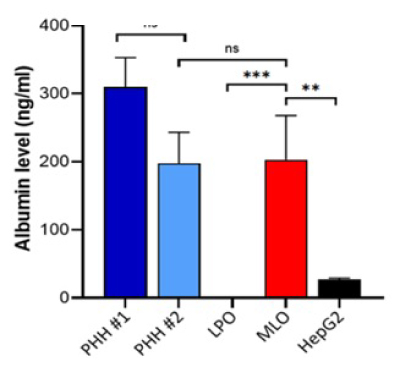
Figure 3.Mature liver organoids (MLO) secrete albumin at levels comparable to primary human hepatocytes (PHH), and significantly higher than liver progenitor organoids (LPO) and HepG2 cells. Two separate lots of primary human hepatocytes (PHH) were tested.
Urea assay
- Organoid seeding
Seed approximately 25-50 LPOs (200-300 μM in diameter) per 10 μL dome into a tissue culture-treated 96-well black plate with flat, clear bottom (for example, Corning 96-Well Black Polystyrene Microplate, CLS3603). - Differentiation
Differentiate LPOs into mature liver organoids following the protocol described above. See workflow above. - Pre-treatment with urea precursors
48-hours prior to performing the urea assay, replace the culture medium with 100 μL of phenol red-free Hepatocyte Culture Medium (Lonza, CC-3198) supplemented with urea production precursors (see Table 3). Incubate for 24-hours.
Note: Use only phenol red-free medium when performing the assay with the Urea Assay Kit III (MAK471). - Ammonium chloride stimulation
After the 24-hour incubation, add 100 μL of Hepatocyte Culture Medium (Lonza, CC-3198) containing 3 mM ammonium chloride (NH4Cl) directly to the wells without removing the existing medium. This results in a final NH4Cl concentration of 1.5 mM. - Sample collection and urea measurement
Incubate for an additional 24-hours, then collect the medium from each well. Perform the urea assay immediately using the Urea Assay Kit III (MAK471) according to the manufacturer’s instructions. - Cell viability assay
Assess cell viability using the CellTiter-Fluor™ Cell Viability Assay (Promega, G6080), following the kit protocol.
Note: Use cell viability data to normalize urea secretion values.

Figure 4.Mature liver organoids secrete urea into the media at a level comparable to primary hepatocytes (PHH) and HepG2 cells. Urea level in medium from mature liver organoids is significantly higher than LPO, indicating functional liver organoids.
CYP1A2, CYP2C9 and CYP3A4 activity assays
- Organoid seeding
Seed approximately 25-50 LPOs (200-300 μM in diameter) per 10 μL dome into a tissue culture-treated 96-well white plate with a flat, clear bottom (for example, Corning 96-Well White Polystyrene Microplate, CLS3610). - Differentiation
Differentiate LPOs into mature liver organoids following the protocol described above. See above workflow. Allocate sufficient wells for each cytochrome P450 (CYP) assay, including conditions for uninduced, induced and induced + inhibitory (rescue) groups. - Induction of CYP activity
To induce CYP activity, replace the medium with Hepatocyte Culture Medium (Lonza, CC-3198) containing specific inducers 48 hours prior to performing the assays:- CYP1A2: 100 μM Omeprazole
- CYP2C9: 25 μM Rifampicin
- CYP3A4: 25 μM Rifampicin
- Incubate organoids with inducer for 24-48 hours. Perform extensive washing with 1X PBS or Basal Plus Medium to remove inducer completely. To completely remove the inducers trapped in the Matrigel®, it is recommended to incubate at 37 °C for 5 minutes between washes.
- CYP activity assays
Measure CYP enzyme activity using the following luciferase-based assays from Promega:- CYP1A2: P450-Glo™ CYP1A2 Induction/Inhibition Assay (Promega, V8421)
- CYP2C9: P450-Glo™ CYP2C9 Assay (Promega, V8791)
- CYP3A4: P450-Glo™ CYP3A4 Assay with Luciferin-IPA (Promega, V9001)
Follow the manufacturer’s instructions for cell-based assays.
- Cell viability assay
Evaluate cell viability using the CellTiter-Glo® 3D Cell Viability Assay Kit (Promega, G9681).
Important: Follow the viability protocol provided within the CYP450 assay kit and not the protocol provided in the CellTiter-Glo® 3D Cell Viability Assay Kit.
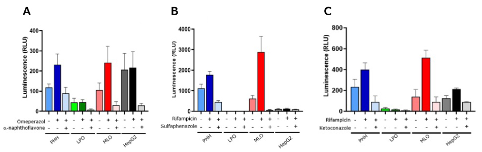
Figure 5.Mature liver organoids (MLO) exhibit both basal and inducible activities of key cytochrome P450 enzymes (A.) CYP1A2, B.) CYP2C9 and C.) CYP3A4) at levels comparable to or exceeding those of primary human hepatocytes (PHH) and HepG2 cells. Enzyme activity can be upregulated by inducers (omeprazole and rifampicin) and suppressed by specific inhibitors (α-naphthoflavone, sulfaphenazole and ketoconazole). In contrast, liver progenitor organoids (LPO) show minimal CYP450 activity.
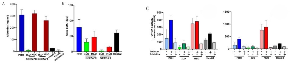
Figure 6.Mature Liver Organoids (MLO) produce higher levels of albumin, urea and CYP3A4 compared to the corresponding Immature Liver Organoids (ILO). A. Comparison of albumin secreted by two representative ILO (SCC570 and SCC571) and their respective MLO; B. Comparison of urea secreted by two representative ILO (SCC570 and SCC571) and their respective MLO; C. Comparison of CYP3A4 activity of two representative ILO (SCC570 graph on the left and SCC571 graph on the right) and their respective MLO. For the CYP3A4 assay, 25 µM rifampicin was used as inducer while 2 µM ketoconazole was used as an inhibitor. Primary human hepatocytes (PHH) were purchased from AnaBios, San Diego, CA. Albumin, urea and CYP3A4 were quantified using Human ALB/ Serum albumin ELISA kit (RAB0603), Urea Assay Kit III (MAK471), and P450-Glo™ CYP3A4 Assay (Promega V9002), respectively.
ALT, AST and GST activity assay
- Organoid seeding
Seed approximately 25-50 LPOs (200-300 μM in diameter) per 10 μL dome into a tissue culture-treated 96-Well black plate with a flat, clear bottom (for example, Corning 96-Well Black Polystyrene Microplate, CLS3603). - Differentiation
Differentiate LPOs into mature liver organoids as described above. See workflow above. - Cell viability assessment
On the day of the assay, perform cell viability measurements using the CellTiter-Fluor™ Cell Viability Assay (Promega, G6080) before the ALT, AST and GST assays. Follow the manufacturer’s instructions.
Note: Viability data will be used to normalize ALT, AST and GST enzyme activity levels. - Membrane permeabilization (optional)
Following the viability assay, treat organoids with 0.2% Triton® X-100 in Basal Plus Medium for 15-20 min at 37 °C to permeabilize cell membranes and release ALT, AST, and GST enzymes into the medium.
Important: This step is applicable only when enzyme activity is measured without drug treatment. If assessing the impact of drug treatments on enzyme activity, do not perform the Triton® X-100 permeabilization step. - Enzyme activity assays
Measure ALT, AST and GST activities using the following assay kits.
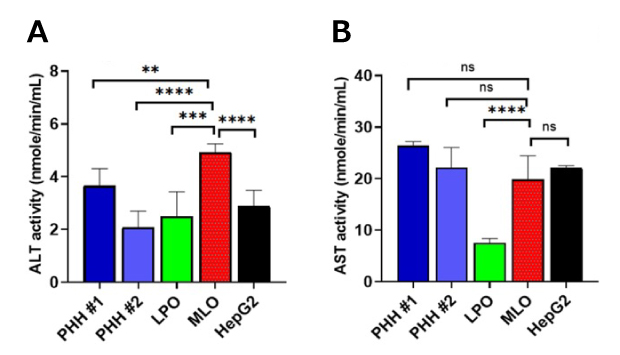
Figure 7.A.)Mature liver organoids possess Alanine Aminotransferase (ALT) and B.) Aspartate Aminotransferase (AST) activities at a level comparable to primary hepatocytes and HepG2 cells, indicating functional liver organoids.