


CB1048 Sigma-AldrichAnti-TNNI2 Rabbit pAb
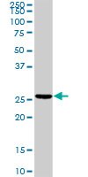

This Anti-TNNI2 Rabbit pAb is validated for use in Immunoblotting for the detection of TNNI2.
More>> This Anti-TNNI2 Rabbit pAb is validated for use in Immunoblotting for the detection of TNNI2. Less<<Anti-TNNI2 Rabbit pAb MSDS (material safety data sheet) or SDS, CoA and CoQ, dossiers, brochures and other available documents.
Synonyms: Anti-Troponin I2
Recommended Products


 Antibody[219217-ALL].jpg)

Overview
| Replacement Information |
|---|
Key Spec Table
| Species Reactivity | Host | Antibody Type |
|---|---|---|
| H, M | Rb | Polyclonal Antibody |
Pricing & Availability
| Catalogue Number | Availability | Packaging | Qty/Pack | Price | Quantity | |
|---|---|---|---|---|---|---|
| CB1048-100UG |
|
100 μg |
|
— |

| References |
|---|
| Product Information | |
|---|---|
| Form | Liquid |
| Formulation | In PBS, pH 7.2. |
| Negative control | 293T cells |
| Positive control | Mouse spleen tissue |
| Preservative | None |
| Quality Level | MQ100 |
| Biological Information | |
|---|---|
| Immunogen | Full-length, human TNNI2 (aa 1-182) |
| Immunogen | Human |
| Host | Rabbit |
| Isotype | IgG |
| Species Reactivity |
|
| Antibody Type | Polyclonal Antibody |
| Physicochemical Information |
|---|
| Dimensions |
|---|
| Materials Information |
|---|
| Toxicological Information |
|---|
| Safety Information according to GHS |
|---|
| Safety Information |
|---|
| Product Usage Statements |
|---|
| Packaging Information |
|---|
| Transport Information |
|---|
| Supplemental Information |
|---|
| Specifications |
|---|
| Global Trade Item Number | |
|---|---|
| Catalogue Number | GTIN |
| CB1048-100UG | 04055977228434 |
Documentation
Anti-TNNI2 Rabbit pAb SDS
| Title |
|---|