Exercise-induced AMPK and pyruvate dehydrogenase regulation is maintained during short-term low-grade inflammation.
Biensø, RS; Olesen, J; van Hauen, L; Meinertz, S; Halling, JF; Gliemann, L; Plomgaard, P; Pilegaard, H
Pflügers Archiv : European journal of physiology
467
341-50
2015
Show Abstract
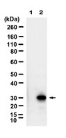
The aim of the present study was to examine the effect of lipopolysaccharide (LPS)-induced inflammation on AMP-activated protein kinase (AMPK) and pyruvate dehydrogenase (PDH) regulation in human skeletal muscle at rest and during exercise. Nine young healthy physically inactive male subjects completed two trials. In an LPS trial, the subjects received a single LPS injection (0.3 ng/kg body weight) and blood samples and vastus lateralis muscle biopsies were obtained before and 2 h after the LPS injection and immediately after a 10-min one-legged knee extensor exercise bout performed approximately 2½ h after the LPS injection. The exercise bout with muscle samples obtained before and immediately after was repeated in a control trial without LPS injection. The plasma tumor necrosis factor α concentration increased 17-fold 2 h after LPS relative to before. Muscle lactate and muscle glycogen were unchanged from before to 2 h after LPS and exercise increased muscle lactate and decreased muscle glycogen in the control (P less than 0.05) and the LPS (0.05 ≤ P less than 0.1) trial with no differences between the trials. AMPK, acetyl-CoA carboxylase (ACC) and PDH phosphorylation as well as PDHa activity were unaffected 2 h after LPS relative to before. Exercise decreased (P less than 0.05) PDH and increased (P less than 0.05) AMPK and ACC phosphorylation as well as increased (P less than 0.05) PDHa activity similarly in the LPS and control trial. In conclusion, LPS-induced inflammation does not affect resting or exercise-induced AMPK and PDH regulation in human skeletal muscle. This suggests that metabolic flexibility during exercise is maintained during short-term low-grade inflammation in humans. | Western Blotting | 24691558
 |
Effect of fatty acids on human bone marrow mesenchymal stem cell energy metabolism and survival.
Fillmore, N; Huqi, A; Jaswal, JS; Mori, J; Paulin, R; Haromy, A; Onay-Besikci, A; Ionescu, L; Thébaud, B; Michelakis, E; Lopaschuk, GD
PloS one
10
e0120257
2015
Show Abstract
Successful stem cell therapy requires the optimal proliferation, engraftment, and differentiation of stem cells into the desired cell lineage of tissues. However, stem cell therapy clinical trials to date have had limited success, suggesting that a better understanding of stem cell biology is needed. This includes a better understanding of stem cell energy metabolism because of the importance of energy metabolism in stem cell proliferation and differentiation. We report here the first direct evidence that human bone marrow mesenchymal stem cell (BMMSC) energy metabolism is highly glycolytic with low rates of mitochondrial oxidative metabolism. The contribution of glycolysis to ATP production is greater than 97% in undifferentiated BMMSCs, while glucose and fatty acid oxidation combined only contribute 3% of ATP production. We also assessed the effect of physiological levels of fatty acids on human BMMSC survival and energy metabolism. We found that the saturated fatty acid palmitate induces BMMSC apoptosis and decreases proliferation, an effect prevented by the unsaturated fatty acid oleate. Interestingly, chronic exposure of human BMMSCs to physiological levels of palmitate (for 24 hr) reduces palmitate oxidation rates. This decrease in palmitate oxidation is prevented by chronic exposure of the BMMSCs to oleate. These results suggest that reducing saturated fatty acid oxidation can decrease human BMMSC proliferation and cause cell death. These results also suggest that saturated fatty acids may be involved in the long-term impairment of BMMSC survival in vivo. | | 25768019
|
Effects of IL-6 on pyruvate dehydrogenase regulation in mouse skeletal muscle.
Biensø, RS; Knudsen, JG; Brandt, N; Pedersen, PA; Pilegaard, H
Pflügers Archiv : European journal of physiology
466
1647-57
2014
Show Abstract
Skeletal muscle regulates substrate choice according to demand and availability and pyruvate dehydrogenase (PDH) is central in this regulation. Circulating interleukin (IL)-6 increases during exercise and IL-6 has been suggested to increase whole body fat oxidation. Furthermore, IL-6 has been reported to increase AMP-activated protein kinase (AMPK) phosphorylation and AMPK suggested to regulate PDHa activity. Together, this suggests that IL-6 may be involved in regulating PDH. The aim of this study was to investigate the effect of a single injection of IL-6 on PDH regulation in skeletal muscle in fed and fasted mice. Fed and 16-18 h fasted mice were injected with either 3 ng · g(-1) recombinant mouse IL-6 or PBS as control. Fasting markedly reduced plasma glucose, muscle glycogen, muscle PDHa activity, as well as increased PDK4 mRNA and protein content in skeletal muscle. IL-6 injection did not affect plasma glucose or muscle glycogen, but increased AMPK and ACC phosphorylation and tended to decrease p38 protein content in skeletal muscle in fasted mice. In addition IL-6 injection reduced PDHa activity in fed mice and increased PDHa activity in fasted mice without significant changes in PDH-E1α phosphorylation or PDP1 and PDK4 mRNA and protein content. The present findings suggest that IL-6 contributes to regulating the PDHa activity and hence carbohydrate oxidation, but the metabolic state of the muscle seems to determine the outcome of this regulation. In addition, AMPK and p38 may contribute to the IL-6-mediated PDH regulation in the fasted state. | | 24221357
|
Xanthene derivatives increase glucose utilization through activation of LKB1-dependent AMP-activated protein kinase.
Kwon, Y; Song, P; Yoon, JH; Ghim, J; Kim, D; Kang, B; Lee, TG; Kim, JA; Choi, JK; Youn, IK; Lee, HK; Ryu, SH
PloS one
9
e108771
2014
Show Abstract
5' AMP-activated protein kinase (AMPK) is a highly conserved serine-threonine kinase that regulates energy expenditure by activating catabolic metabolism and suppressing anabolic pathways to increase cellular energy levels. Therefore AMPK activators are considered to be drug targets for treatment of metabolic diseases such as diabetes mellitus. To identify novel AMPK activators, we screened xanthene derivatives. We determined that the AMPK activators 9H-xanthene-9-carboxylic acid {2,2,2-trichloro-1-[3-(3-nitro-phenyl)-thioureido]-ethyl}-amide (Xn) and 9H-xanthene-9-carboxylic acid {2,2,2-trichloro-1-[3-(3-cyano-phenyl)-thioureido]-ethyl}-amide (Xc) elevated glucose uptake in L6 myotubes by stimulating translocation of glucose transporter type 4 (GLUT4). Treatment with the chemical AMPK inhibitor compound C and infection with dominant-negative AMPKa2-virus inhibited AMPK phosphorylation and glucose uptake in myotubes induced by either Xn or Xc. Of the two major upstream kinases of AMPK, we found that Xn and Xc showed LKB1 dependency by knockdown of STK11, an ortholog of human LKB1. Single intravenous administration of Xn and Xc to high-fat diet-induced diabetic mice stimulated AMPK phosphorylation of skeletal muscle and improved glucose tolerance. Taken together, these results suggest that Xn and Xc regulate glucose homeostasis through LKB1-dependent AMPK activation and that the compounds are potential candidate drugs for the treatment of type 2 diabetes mellitus. | | 25250787
|
AAV8-mediated Sirt1 gene transfer to the liver prevents high carbohydrate diet-induced nonalcoholic fatty liver disease.
Vilà, L; Elias, I; Roca, C; Ribera, A; Ferré, T; Casellas, A; Lage, R; Franckhauser, S; Bosch, F
Molecular therapy. Methods & clinical development
1
14039
2014
Show Abstract
Nonalcoholic fatty liver disease (NAFLD) is the most common hepatic disease worldwide, and evidence suggests that it promotes insulin resistance and type 2 diabetes. Caloric restriction (CR) is the only available strategy for NAFLD treatment. The protein deacetylase Sirtuin1 (SIRT1), which is activated by CR, increases catabolic metabolism and decreases lipogenesis and inflammation, both involved in the development of NAFLD. Here we show that adeno-associated viral vectors of serotype 8 (AAV8)-mediated liver-specific Sirt1 gene transfer prevents the development of NAFLD induced by a high carbohydrate (HC) diet. Long-term hepatic SIRT1 overexpression led to upregulation of key hepatic genes involved in β-oxidation, prevented HC diet-induced lipid accumulation and reduced liver inflammation. AAV8-Sirt1-treated mice showed improved insulin sensitivity, increased oxidative capacity in skeletal muscle and reduced white adipose tissue inflammation. Moreover, HC feeding induced leptin resistance, which was also attenuated in AAV8-Sirt1-treated mice. Therefore, AAV-mediated gene transfer to overexpress SIRT1 specifically in the liver may represent a new gene therapy strategy to counteract NAFLD and related diseases such as type 2 diabetes. | | 26015978
|
Increased postexercise insulin sensitivity is accompanied by increased AS160 phosphorylation in slow-twitch soleus muscle.
Iwabe, M; Kawamoto, E; Koshinaka, K; Kawanaka, K
Physiological reports
2
2014
Show Abstract
A single bout of exercise can enhance insulin-stimulated glucose uptake in both fast-twitch (type II) and slow-twitch (type I) skeletal muscle for several hours postexercise. Akt substrate of 160 kDa (AS160) is most distal insulin signaling proteins that have been proposed to contribute to the postexercise enhancement of insulin action in fast-twitch muscle. In this study, we examined whether the postexercise increase in insulin action of glucose uptake in slow-twitch muscle is accompanied by increased phosphorylation of AS160 and its paralog TBC1D1. Male Wistar rats (~1-month-old) were exercised on a treadmill for 180 min (9 m/min). Insulin (50 μU/mL)-stimulated glucose uptake was increased at 2 h after cessation of exercise in soleus muscle composed of predominantly slow-twitch fibers. This postexercise increase in insulin action of glucose uptake was accompanied by increased phosphorylation of AS160 (detected by phospho-Thr642 and phospho-Ser588 antibody). On the other hand, prior exercise did not increase phosphorylation of TBC1D1 (detected by phospho-Thr590) at 2 h postexercise. These results suggest the possibility that an enhancement in AS160 phosphorylation but not TBC1D1 phosphorylation is involved with increased postexercise insulin action of glucose uptake in slow-twitch muscle. | | 25501433
|
Two weeks of metformin treatment enhances mitochondrial respiration in skeletal muscle of AMPK kinase dead but not wild type mice.
Kristensen, JM; Larsen, S; Helge, JW; Dela, F; Wojtaszewski, JF
PloS one
8
e53533
2013
Show Abstract
Metformin is used as an anti-diabetic drug. Metformin ameliorates insulin resistance by improving insulin sensitivity in liver and skeletal muscle. Reduced mitochondrial content has been reported in type 2 diabetic muscles and it may contribute to decreased insulin sensitivity characteristic for diabetic muscles. The molecular mechanism behind the effect of metformin is not fully clarified but inhibition of complex I in the mitochondria and also activation of the 5'AMP activated protein kinase (AMPK) has been reported in muscle. Furthermore, both AMPK activation and metformin treatment have been associated with stimulation of mitochondrial function and biogenesis. However, a causal relationship in skeletal muscle has not been investigated. We hypothesized that potential effects of in vivo metformin treatment on mitochondrial function and protein expressions in skeletal muscle are dependent upon AMPK signaling. We investigated this by two weeks of oral metformin treatment of muscle specific kinase dead α(2) (KD) AMPK mice and wild type (WT) littermates. We measured mitochondrial respiration and protein activity and expressions of key enzymes involved in mitochondrial carbohydrate and fat metabolism and oxidative phosphorylation. Mitochondrial respiration, HAD and CS activity, PDH and complex I-V and cytochrome c protein expression were all reduced in AMPK KD compared to WT tibialis anterior muscles. Surprisingly, metformin treatment only enhanced respiration in AMPK KD mice and thereby rescued the respiration defect compared to the WT mice. Metformin did not influence protein activities or expressions in either WT or AMPK KD mice.We conclude that two weeks of in vivo metformin treatment enhances mitochondrial respiration in the mitochondrial deficient AMPK KD but not WT mice. The improvement seems to be unrelated to AMPK, and does not involve changes in key mitochondrial proteins. | | 23341947
|
Activation of sterol regulatory element binding protein and NLRP3 inflammasome in atherosclerotic lesion development in diabetic pigs.
Li, Y; Xu, S; Jiang, B; Cohen, RA; Zang, M
PloS one
8
e67532
2013
Show Abstract
Aberrantly elevated sterol regulatory element binding protein (SREBP), the lipogenic transcription factor, contributes to the development of fatty liver and insulin resistance in animals. Our recent studies have discovered that AMP-activated protein kinase (AMPK) phosphorylates SREBP at Ser-327 and inhibits its activity, represses SREBP-dependent lipogenesis, and thereby ameliorates hepatic steatosis and atherosclerosis in insulin-resistant LDLR(-/-) mice. Chronic inflammation and activation of NLRP3 inflammasome have been implicated in atherosclerosis and fatty liver disease. However, whether SREBP is involved in vascular lipid accumulation and inflammation in atherosclerosis remains largely unknown.The preclinical study with aortic pouch biopsy specimens from humans with atherosclerosis and diabetes shows intense immunostaining for SREBP-1 and the inflammatory marker VCAM-1 in atherosclerotic plaques. The cleavage processing of SREBP-1 and -2 and expression of their target genes are increased in the well-established porcine model of diabetes and atherosclerosis, which develops human-like, complex atherosclerotic plaques. Immunostaining analysis indicates an elevation in SREBP-1 that is primarily localized in endothelial cells and in infiltrated macrophages within fatty streaks, fibrous caps with necrotic cores, and cholesterol crystals in advanced lesions. Moreover, concomitant suppression of NAD-dependent deacetylase SIRT1 and AMPK is observed in atherosclerotic pigs, which leads to the proteolytic activation of SREBP-1 by diminishing the deacetylation and Ser-372 phosphorylation of SREBP-1. Aberrantly elevated NLRP3 inflammasome markers are evidenced by increased expression of inflammasome components including NLPR3, ASC, and IL-1β. The increase in SREBP-1 activity and IL-1β production in lesions is associated with vascular inflammation and endothelial dysfunction in atherosclerotic pig aorta, as demonstrated by the induction of NF-κB, VCAM-1, iNOS, and COX-2, as well as by the repression of eNOS.These translational studies provide in vivo evidence that the dysregulation of SIRT1-AMPK-SREBP and stimulation of NLRP3 inflammasome may contribute to vascular lipid deposition and inflammation in atherosclerosis. | | 23825667
|
Adiponectin attenuates angiotensin II-induced oxidative stress in renal tubular cells through AMPK and cAMP-Epac signal transduction pathways.
Fang, F; Liu, GC; Kim, C; Yassa, R; Zhou, J; Scholey, JW
American journal of physiology. Renal physiology
304
F1366-74
2013
Show Abstract
Obesity is a risk factor for chronic kidney disease (CKD) progression. Circulating levels of adiponectin, an adipokine, decrease with obesity and play a protective role in the cardiovascular system. We hypothesized that adiponectin might also protect the kidney. Because activation of the renin-angiotensin system (RAS) is a contributor to CKD progression, we tested our hypothesis by studying the interactions between adiponectin and angiotensin II (ANG II) in renal tubular cells. Primary human renal proximal tubule cells expressed both adiponectin receptor 1 and 2 (adipoR1 and R2). ANG II-induced NADPH oxidase activation and oxidative stress were attenuated by adiponectin and dependent on adipoR1. Activation of AMPK with 5-aminoimidazole-4-carboxamide-1-β-d-ribofuranoside (AICAR) mimicked, while inhibition of AMPK with compound C abrogated, the effect of adiponectin on ANG II-induced activation of NADPH oxidase. Similarly, the effect of adiponectin was recapitulated by the stable cAMP analogs 4-chlorophenylthio (pCPT)-cAMP and dibutyryl (db)-cAMP and blocked by the adenylate cyclase inhibitor SQ22536. Adiponectin did not activate PKA in renal tubular cells, and the specific PKA inhibitor myristoylated PKI (14-22) amide failed to block the inhibitory effect of adiponectin on ANG II-induced NADPH oxidase activation. In contrast, the specific Epac activator 8-(4-chlorophenylthio)-2'-O-methyl (8-CPT-2-O-Me)-cAMP blocked ANG II-induced activation of NADPH oxidase, an effect that was reversed by coincubation with the AMPK inhibitor compound C. Finally, adiponectin attenuated ANG II-induced NF-κB activation and fibronectin protein expression. These in vitro findings support the hypothesis that adiponectin may attenuate the deleterious effects of ANG II in the kidney and play a protective role in CKD. | | 23535586
|
Resistance to aerobic exercise training causes metabolic dysfunction and reveals novel exercise-regulated signaling networks.
Lessard, SJ; Rivas, DA; Alves-Wagner, AB; Hirshman, MF; Gallagher, IJ; Constantin-Teodosiu, D; Atkins, R; Greenhaff, PL; Qi, NR; Gustafsson, T; Fielding, RA; Timmons, JA; Britton, SL; Koch, LG; Goodyear, LJ
Diabetes
62
2717-27
2013
Show Abstract
Low aerobic exercise capacity is a risk factor for diabetes and a strong predictor of mortality, yet some individuals are "exercise-resistant" and unable to improve exercise capacity through exercise training. To test the hypothesis that resistance to aerobic exercise training underlies metabolic disease risk, we used selective breeding for 15 generations to develop rat models of low and high aerobic response to training. Before exercise training, rats selected as low and high responders had similar exercise capacities. However, after 8 weeks of treadmill training, low responders failed to improve their exercise capacity, whereas high responders improved by 54%. Remarkably, low responders to aerobic training exhibited pronounced metabolic dysfunction characterized by insulin resistance and increased adiposity, demonstrating that the exercise-resistant phenotype segregates with disease risk. Low responders had impaired exercise-induced angiogenesis in muscle; however, mitochondrial capacity was intact and increased normally with exercise training, demonstrating that mitochondria are not limiting for aerobic adaptation or responsible for metabolic dysfunction in low responders. Low responders had increased stress/inflammatory signaling and altered transforming growth factor-β signaling, characterized by hyperphosphorylation of a novel exercise-regulated phosphorylation site on SMAD2. Using this powerful biological model system, we have discovered key pathways for low exercise training response that may represent novel targets for the treatment of metabolic disease. | | 23610057
|