Chiudendo, la configurazione personalizzata non sarà salvata, a meno che l'articolo non sia stato aggiunto al carrello della spesa o ai prodotti preferiti.
Clicchi OK per chiudere MILLIPLEX® MAP, oppure Annulla per tornare alla selezione.
Scelga pannelli su misura e kit premiscelati, OPPURE le microsfere MAPmates™ per la trasduzione del segnale
Progetti e calcoli il prezzo dei Suoi kit MILLIPLEX® MAP.
Pannelli su misura e kit premiscelati
La nostra ampia gamma comprende pannelli multiplex che consentono di scegliere, nell'ambito del pannello, gli analiti più adatti per le Sue esigenze. Oppure Lei può scegliere le citochine premiscelate o i kit single plex.
Kit e MAPmates™ per studi di trasduzione del segnale
Scelga i kit premiscelati che permettono di esplorare interi processi e pathway. Oppure progetti kit su misura scegliendo le microsfere MAPmates™ single plex e seguendo le linee guida fornite.
Le seguenti MAPmates™ non possono essere dosate insieme: -MAPmates™ che richiedono tamponi differenti per l'analisi. -MAPmate™ fosfo-specifiche e totali, es GSK3β totale e GSK3β (Ser 9). -MAPmates™ PanTyr e sito-specifiche come fosfo-recettore EGF e fosfo-STAT1 (Tyr701). -Più di 1 fosfo-MAPmate™ per un solo bersaglio (Akt, STAT3). -GAPDH e β-Tubulina non possono essere miscelati con kit o MAPmates™ contenenti panTyr.
.
Numero di catalogo
Descrizione dell'ordine
Qtà/conf
Elenca
Questo articolo è stato aggiunto ai prodotti preferiti.
Selezioni una specie, un tipo di pannello, un kit o un tipo di campione
Per cominciare a progettare il Suo kit MILLIPLEX® MAP, selezioni una specie, un tipo di pannello o un kit di Suo interesse.
Custom Premix Selecting "Custom Premix" option means that all of the beads you have chosen will be premixed in manufacturing before the kit is sent to you.
Catalogue Number
Ordering Description
Qty/Pack
List
Questo articolo è stato aggiunto ai prodotti preferiti.
Specie
Tipo di pannello
Kit selezionato
Qtà
Numero di catalogo
Descrizione dell'ordine
Qtà/conf
Prezzo di listino
96-Well Plate
Qtà
Numero di catalogo
Descrizione dell'ordine
Qtà/conf
Prezzo di listino
Aggiungi altri reagenti (Le microsfere MAPmate devono essere utilizzate con un tampone ed un kit di rilevazione)
Qtà
Numero di catalogo
Descrizione dell'ordine
Qtà/conf
Prezzo di listino
48-602MAG
Buffer Detection Kit for Magnetic Beads
1 Kit
Opzione salva-spazio I Clienti che ordinano diversi kit possono scegliere di ridurre lo spazio necessario per lo stoccaggio, rinunciando al confezionamento del kit e ricevendo i vari reagenti per il saggio multiplex in buste di plastica.
Questo articolo è stato aggiunto ai prodotti preferiti.
Il prodotto è stato aggiunto al carrello.
Ora può disegnare un altro kit su misura, scegliere un kit premiscelato, procedere con il check out dell'ordine, oppure chiudere lo strumento per gli ordini on-line.
The goal of drug discovery is to find well-qualified candidate drugs expected to provide therapeutic benefit. Use of relevant and information-rich assays can rapidly eliminate candidates that either do not affect the appropriate response or cause some unforeseen lethality, and can save drug companies time and money that is often wasted pursuing clinical trials with flawed drug candidates. Many of the Amnis® imaging flow cytometry's applications are ideally suited for characterizing drug candidates, including cell signaling, cell-cell interactions, chemokine-induced shape change, cell death, co-localization, and phagocytosis, to name a few. Furthermore, these applications work with primary cells such as whole blood leukocytes and can often be multiplexed.
Co-Localization of an Antibody-Drug Conjugate to Endosomes or Lysosomes
In this experiment we use the capabilities of the Imagestream®x system to measure the transit of a fluorescently labeled antibody-drug conjugate (ADC) through the cellular endocytic pathway. The high spatial resolution afforded by the Imagestream®x system allowed accurate measurement of ADC co-localization to endosomes and lysosomes. See the application note for more details.
Barcoding
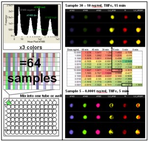
Screening drug candidates requires the rapid analysis of large numbers of samples. Multiplexing samples on the Imagestream®x by mixing dozens of samples from a time course or dose response titration into one tube dramatically increases the sample throughput of the system. This is achieved through fluorescent cell barcoding with subsequent identification of each sample based on a pattern of fluorescent dye and staining intensity unique to each treatment/time point. Automated location of populations based on fluorescence intensity and color allows deconvolution of the barcoded samples after acquisition.
Here is an example of measuring the movement of NF-κB from the cytoplasm to the nucleus in response to varying doses and times of exposure to TNF-α. By labeling 64 separate samples with different combinations of 3 dyes (AlexaFluor 405, 488, and 660) at 4 intensity levels (43 = 64) it was possible to run one tube of the combined samples on the Imagestream®x and acquire a single file. After acquisition of over 600,000 images in 10-15 minutes, the color and intensity combinations of the samples were deconvoluted for further quantitative image analysis. Mean Similarity scores are reported in the spreadsheet and a heatmap has been applied. See the cell signaling application for more information about the translocation assay. The combination of high speed image acquisition and automated quantitative image analysis provided by the Imagestream®x system offers a major advancement in the field of drug discovery.