Assess Efficacy and Selectivity of a Bispecific Antibody Using Organoid-Based Co-Cultures
Therapeutic and novel drug development starts with compound discovery, but once a lead compound has been identified researchers need to evaluate the specificity, efficacy, and mechanism of action. Here we describe how organoid co-cultures with T cells can form a powerful preclinical platform to evaluate the efficacy, specificity, and mechanism of action of bispecific antibodies.
The preclinical PDO-T cell co-culture platform
For this collaboration, we utilized a lead candidate from the client, a bispecific T cell engager (BiTE) for solid tumor indications that required a preclinical system capable of accurately assessing both efficacy and safety of this candidate. Conventional 2D models lacked the complexity that was needed to accurately evaluate the tumor-selective killing and immune engagement of the BiTE within a human-relevant microenvironment. Specifically, a translational platform was needed that could preserve tumor heterogeneity, reflect patient-specific antigen expression, and support functional co-culture with T cells to de-risk downstream in vivo studies and guide candidate optimization.
We applied our PDO–T cell co-culture platform to model antigen-specific immune engagement and cytotoxicity. Using matched tumor and normal non-small cell lung cancer (NSCLC) patient-derived organoids (PDO), we established co-cultures with primary T cells to assess the bispecific antibody’s ability to induce selective killing towards tumor cells and activate T cells. The system enabled head-to-head comparison of on-target efficacy and potential off-tumor toxicity, with quantitative functional readouts – providing the client with translationally predictive data ahead of in vivo studies.
Our approach
We designed an experiment with three key assays:
- Target antigen profiling: Flow cytometry was used to confirm tumor-associated antigen (TAA) expression across tumor and matched normal PDOs.
- Cytotoxicity assay: Cell death in tumor and normal PDOs was measured following treatment with the bispecific antibody in the presence or absence of T cells.
- T cell activation: IFN-γ secretion was quantified as a proxy for T cell activation upon engagement with the bispecific antibody.
PDO-T cell co-culture results
Organoid model selection
Leveraging our large living biobank, we suggested testing the expression of the TAA in NSCLC tumor organoids and matched normal lung organoids. Using immunohistochemistry (IHC) (Figure 1A) and flow cytometry (Figure 1B) data, we supported our client in selecting the most relevant organoid models.
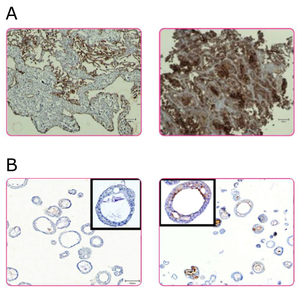
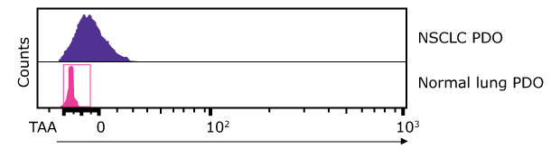
Figure 1.A. Representative images of IHC staining for the TAA in human tumor tissue and matched normal epithelium and corresponding patient-derived organoids. B. White arrowheads indicate expression of the TTA in tumor epithelial cells. Flow cytometry analysis of TAA in NSCLC PDO and matched normal lung PDO.
Differential target expression
Through ICH and flow cytometry we confirmed expression of the TAA specifically in the tumor PDOs. This ensured both specificity and the relevance of the chosen models for subsequent efficacy and toxicity testing.
T cell–dependent, tumor-selective cytotoxicity
The bispecific antibody (BiTE) induced significant killing of tumor PDOs when co-cultured with T cells, while normal PDOs were largely spared under identical conditions (Figure 2). No cell death occurred in the absence of T cells or when using a non-targeting BiTE (nonspecific), confirming both antigen specificity and T cell dependence.
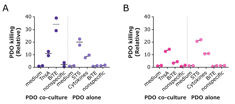
Figure 2.Day 2 quantification of cell death fluorescent signal in A. tumor and B. normal PDOs when exposed to a BiTE directed to a tumor-specific antigen. For organoid killing, staurosporine (STS) and cytokines are used as positive control and medium only as negative control. T cell Trans activator is included as a control for T-cell activation (TnsA). In absence of T- cells, the tested BiTE, TnsA, and nonspecific ab are not able to induce PDO cell death.
Discover how we accelerated the development of a bispecific antibody within 5 years!
Functional T cell activation
IFN-γ secretion was significantly elevated in co-cultures treated with the targeted BiTE, indicating effective activation of T cells in response to TAA recognition on tumor PDOs.
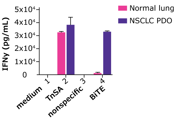
Figure 3.IFN-γ levels secreted by T cells in co-culture with PDOs and in presence of targeted BiTE and a non-specific BiTE as control. Transactivator (TnsA) was included as positive control to induce IFN-γ secretion.
PDO-T cell co-culture system key takeaways
This study demonstrated that the PDO–T cell co-culture system provided insight into the efficacy and selectivity of the bispecific antibody tested, supporting the progression of the bispecific antibody toward in vivo validation. In the study, the bispecific antibody elicited antigen-specific, T cell–dependent killing of NSCLC PDOs with minimal off-tumor effects on matched normal organoids. The bispecific antibody also induced elevated IFN-γ secretion, confirming functional T cell engagement.
For bispecific antibody developers, this platform offers a translationally relevant, human-derived system to de-risk immunotherapeutic candidates early in development by modeling tumor–immune dynamics and off-tumor toxicity in vitro.
Looking to use HUB Organoids® for your drug development program? Speak to an expert now!
Related products