Modeling human fibroblast-epithelial interactions with patient-derived organoids
Organoids to identify IBD targets
Inflammatory bowel disease (IBD) is the term used to describe chronic conditions of inflammation and swelling in the intestine; the most common forms of IBD are ulcerative colitis and Crohn’s disease. Treatment for IBD usually includes either surgery or medication, with the goal of reducing the inflammation that causes the symptoms.
Due to poor translational relevance of cell cultures and the inability of animal models to reflect patient IBD insights pathogenesis, we helped develop a novel 3D organoid co-culture system that recapitulated the interaction between the epithelium and the surrounding fibroblasts some of the disease hallmarks. A role for proinflammatory fibroblast in the pathogenesis of IBD has been suggested but novel therapeutic interventions targeting the epithelium-fibroblast axis have never been tested. Our 3D organoid co-cultures better recapitulate key IBD hallmarks, helping researchers understand fibroblast-epithelial cell interactions and how the fibroblast pathways fuel IBD inflammation and facilitating drug discovery and development.
Here we describe how we modeled human fibroblast-epithelial interactions using organoids to identify novel IBD targets.
3D organoid co-culture assay overview
We proposed a physiologically relevant 3D co-culture assay, combining intestinal fibroblasts (± inflammatory stimulation) with IBD patient-derived organoids to enable dynamic signaling and direct interaction in a 3D format compatible with different readouts. Our co-culture set-up aimed to quantify the inflammatory state, epithelial cell death and assess the proliferative or differentiated state of the epithelium. We verified our assay using standard of care, following in-depth optimizations of all readouts and inflammation triggers, providing the client with a valuable tool for their preclinical IBD drug discovery and development.
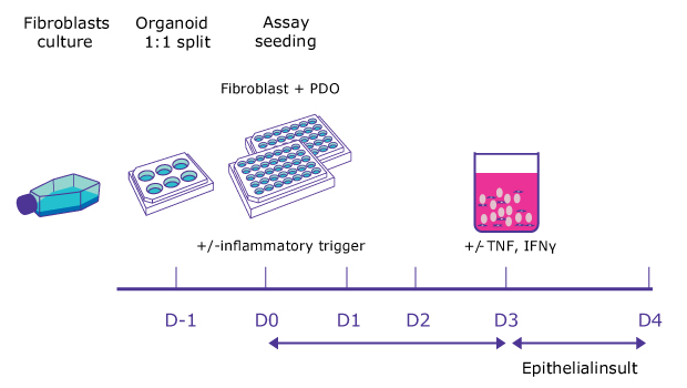
Figure 1.Schematic of co-culturing human fibroblasts with IBD patient-derived organoids study design. The study protocol included induction of an inflammatory trigger for an inflammatory fibroblast phenotype followed by a cytokine-induced epithelial damage mimicking mucosal IBD environment. In this phase, along with the client, we did an intensive literature search to understand probable readouts from the experiment that could replicate the hallmarks of IBD pathogenesis.
Establishing an organoid-fibroblast co-culture platform
Establishing a reliable and reproducible in vitro model is a critical first step and involved optimizing the 3D co-culture conditions for fibroblasts and IBD patient-derived epithelial organoids. We carefully identified and titrated the concentration of an inflammatory trigger to induce the desired inflammatory fibroblast phenotype and performed multiple rounds of culture condition optimization to ensure the system’s robustness.
Organoid swelling served as the key readout to visualize the impact of the inflammatory fibroblasts on the epithelium. Importantly, the reproducibility of this swelling metric was rigorously assessed with a Z’ factor of >0.5 (Figure 2), demonstrating excellent assay quality and reliability for subsequent experiments
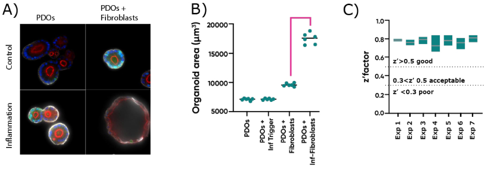
Figure 2.A) Representative confocal microscopy images of PDOs cultured alone or with fibroblasts with or without inflammatory stimuli. PDOs were stained for Phalloidin (red), KI67 (green), KRT20 (white) and DAPI (blue). B) Organoid area change in response to the presence of fibroblasts and/or inflammatory stimuli. C) Z’ scores for organoid area change of co-cultures exposed to inflammation across experiments. Each experiment included 3 plates with independent controls.
Characterizing inflammatory fibroblast and epithelial cell crosstalk
One of our core objectives was to characterize the complex communication between inflammatory fibroblasts and epithelial cells within the co-culture. To that end, we investigated the role of various soluble mediators secreted by fibroblasts and epithelial cells in response to the inflammatory stimulus. Our data revealed that the inflammatory trigger significantly changed the fibroblast’s phenotype and that the co-culture allows for permanent cross-communication emphasizing the effect of the inflammation (Figure 3).
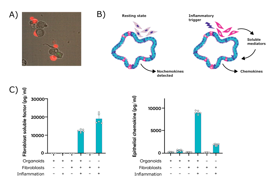
Figure 3.A) Fibroblasts (red) associate with the intestinal epithelium when co-cultured with PDOs. B) Cartoon representing the crosstalk between (proinflammatory) fibroblasts and epithelial cells. C) Inflammatory trigger drives fibroblasts to activate chemokine production on epithelial cells.
Recapitulating IBD hallmarks
In addition to soluble mediator analysis, we examined other key hallmarks of IBD within the co-culture system. Notably, we observed that inflammatory stimuli resulted in a significant decrease in organoid cell proliferation only when the inflammatory fibroblasts were present, mirroring the impaired tissue regeneration often seen in IBD.
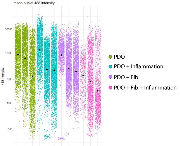
Figure 4.Mean EdU-488 nuclear intensity quantification demonstrates daily decline in cell proliferation when PDOs are co-cultured with fibroblast in presence of a pro-inflammatory stimulus. Each datapoint represents a single nucleus.
Co-culture assay validation
To further validate the translational relevance of our co-culture assay, we assessed the effect of a standard-of-care treatment for IBD. The addition of tofacitinib, a clinically relevant therapeutic, resulted in a reduction of both organoid swelling and cell death when exposed to damage-inducing cytokines, demonstrating the assay’s utility for evaluating the potential of new therapeutic candidates.
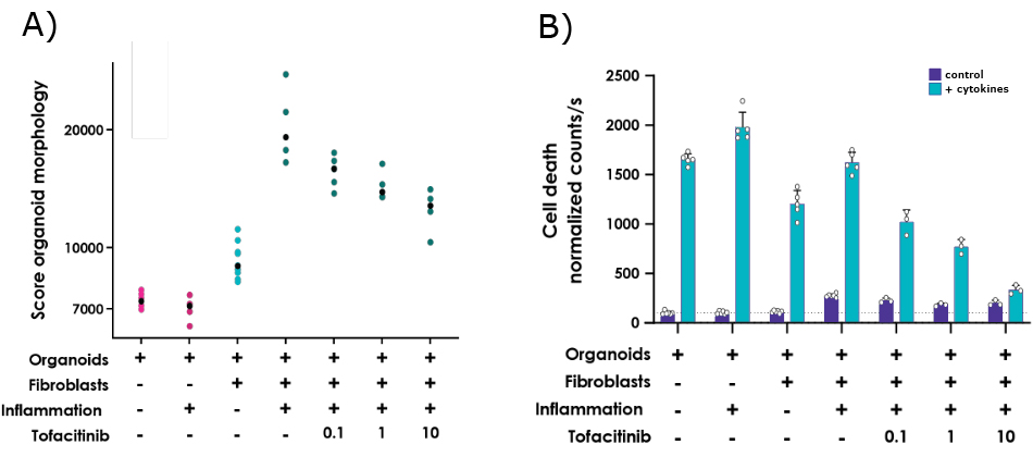
Figure 5.A) Tofacitinib limits the organoid swelling and B) cytokine-induced cell death induced by the proinflammatory fibroblasts.
Interested in patient-derived organoids for IBD? Contact an expert now!