Membrane Immunodetection Devices

Western blotting immunodetection involves the use of antibodies to detect target protein. Blotted membranes containing transferred proteins are blocked, followed by antibody incubation and detection using chemiluminescence, colorimetric, or fluorescent methods. Membrane immunodetection devices facilitate the processing of Western blots by simplifying workflows and accelerating detection times.
Section Overview
Products

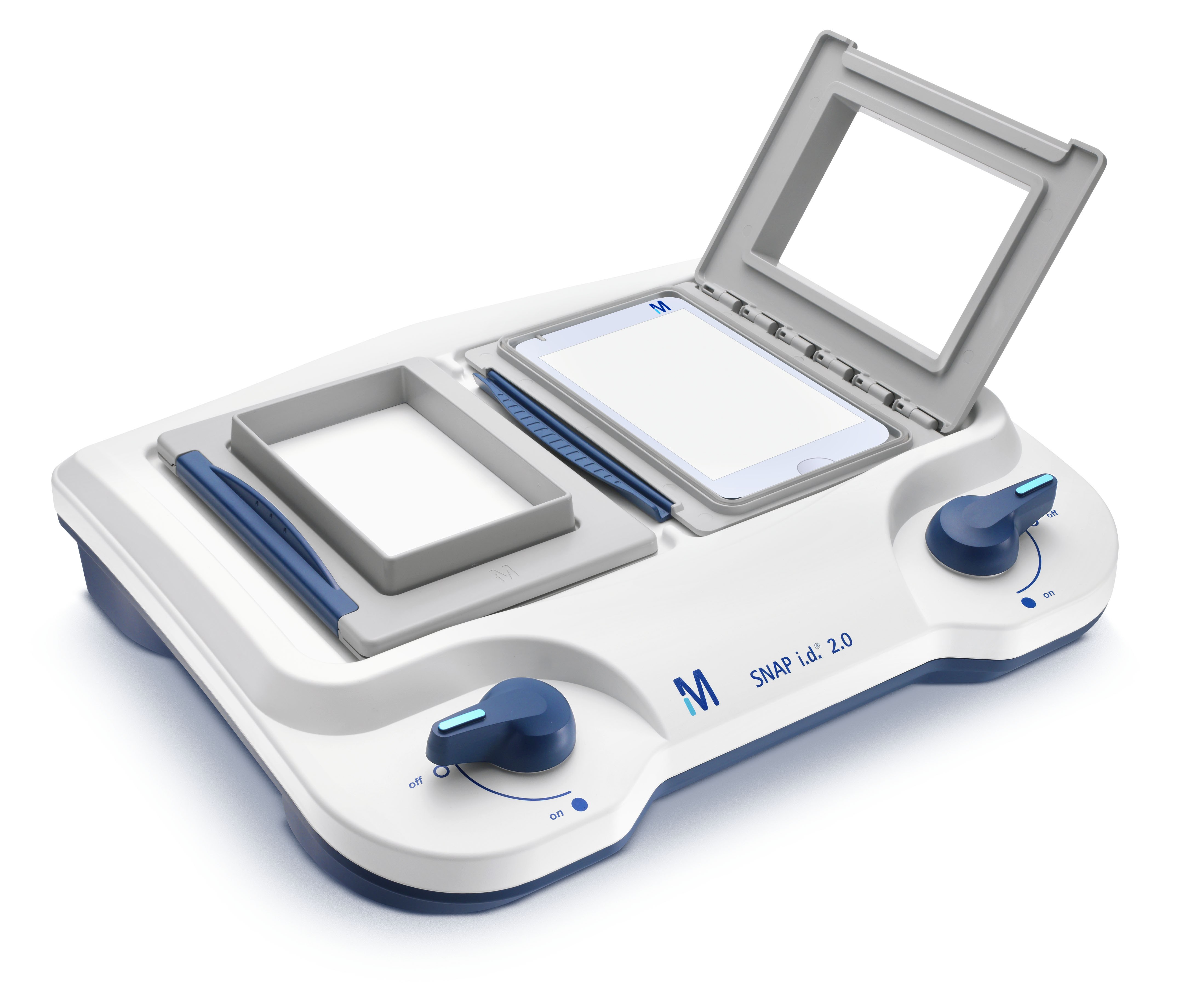
SNAP i.d.® 2.0 Protein Detection System
The SNAP i.d.® 2.0 Protein Detection System is a unique, vacuum-driven system that pulls reagents through the membrane, increasing membrane contact while eliminating dependence on diffusion or agitation. This improvement in three-dimensional reagent distribution decreases the length of time required for immunodetection. While traditional Western blotting requires 4 to 24 hours for the blocking, antibody incubation, and washing steps, the SNAP i.d.® 2.0 protocol takes only 30 minutes with no loss of signal intensity or reduction in blot quality. All immunodetection steps after protein transfer to a membrane (i.e., blocking, washing, and primary and secondary antibody incubations) can be performed with the SNAP i.d.® 2.0 System.
Related Resources
- Handbook: Protein Blotting Tips and Tricks
This handbook represents the collective experience of our application scientists, who are actively engaged in advancing the science of protein blotting and detection.
- Article: 30-minute Immunodetection Protocol Using the SNAP i.d.® 2.0 System
The SNAP i.d. 2.0 Protein Detection System is the second generation of the SNAP i.d. method for detecting immunoreactive proteins on Western blots.
- Brochure: Rethink Western Blotting
Access to our Western blotting expertise is easy—flip to our troubleshooting section at the end of this brochure, or learn more.
To continue reading please sign in or create an account.
Don't Have An Account?