Measurement of proteins such as cytokines, antibodies, and growth factors is essential for elucidating cell signaling pathways, uncovering disease mechanisms, identifying and validating targets for drug discovery, characterizing immune response, and evaluating drug pharmacokinetics and pharmacodynamics. Several methods for qualitative and quantitative protein detection exist, including ELISA (Enzyme-Linked Immunosorbent Assay) and ELISpot (Enzyme-Linked ImmunoSpot). While both techniques rely on antibodies and enzyme-driven reactions for detection, they serve different experimental goals. Here, we compare key principles, applications, strengths, and limitations of ELISA and ELISpot to help you select the right experimental method for your studies.
Section Overview
- What is an ELISA?
- How Does ELISA Work?
- What is ELISpot?
- How Does ELISpot Work?
- Choosing between ELISA and ELISpot
- Using ELISA and ELISpot Together
- Related Products
What is an ELISA?
ELISA (Enzyme-Linked Immunosorbent Assay) is one of the most widely used immunoassay platforms for detecting and quantitating amounts of soluble proteins in samples including sera, plasma, and cell culture supernatants. ELISA works through capture of target analyte using specific antibodies and generation of a measurable signal - typically colorimetric or luminescent - that correlates with the concentration of the analyte.
There are four main types of ELISA:
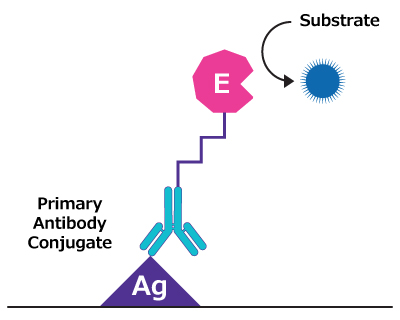
- Direct ELISA employs a primary antibody that is enzyme-labeled (e.g., HRP or AP conjugate). This format offers a straightforward and fast protocol, but typically has lower sensitivity due to limited signal amplification.
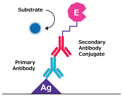
- Indirect ELISA enhances sensitivity by using a two-step detection system involving a primary antibody that binds to target protein and an enzyme-labeled secondary antibody. This format allows for signal amplification, but may introduce cross-reactivity when using an anti-species secondary antibody.

- Sandwich ELISA is highly specific and suitable for detecting proteins and analytes in complex samples. This assay format relies on two antibodies: a “capture” antibody coated to the microplate well that immobilizes target protein, and a “detection” antibody that binds a different epitope on the same protein to create a “sandwich”. While sandwich ELISA requires well-matched antibody pairs, its high specificity and sensitivity make it a preferred method when quantitating low-abundance analytes in complex matrices.
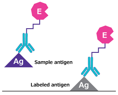
- Competitive ELISA is best suited for small molecules or antigens with limited epitopes or limited availability of antibodies. In this format, antigen in your sample competes with a labeled antigen for antibody binding. Competitive ELISA provides high sensitivity but involves a more intricate setup and inverse signal readout (i.e., signal decreases with increasing antigen concentration).
Each ELISA format has its own strengths. Choosing the right format depends on your experimental needs and available reagents.
How Does ELISA Work?
In a sandwich ELISA, a high-binding microplate is coated with capture antibody specific to the target protein. After blocking to prevent non-specific binding, the sample is added and incubated to allow the target protein to bind to capture antibody. A biotinylated detection antibody is then introduced, binding to a different epitope on the same protein. This is followed by addition of a streptavidin-enzyme conjugate (e.g., streptavidin-HPR, streptavidin-AP). A colorimetric or chemiluminescent substrate for the enzyme is then added to produce a signal proportional to the amount of protein present. The signal is measured using a plate reader. Protein concentrations are typically determined using a standard curve that plots optical density (OD) values or relative light units (RLUs) against known concentrations of the target analyte. This curve is used to interpolate the concentrations of unknown samples.
What is ELISpot?
ELISpot (Enzyme-Linked ImmunoSpot) is a highly sensitive immunoassay for counting cells that secrete specific protein(s) - typically cytokines or antibodies. Unlike ELISA, which measures the overall concentration of analytes in a fluid sample, ELISpot provides a detailed snapshot of cellular response at the single-cell level. This is particularly valuable in immunology research, where detecting rare antigen-specific T or B cells is critical. ELISpot assays are frequently used in vaccine development, autoimmune studies, and cancer immunotherapy monitoring.

Figure 1.ELISpot workflow.
How Does ELISpot Work?
In ELISpot, cells are added to a PVDF filter plate pre-coated with a capture antibody specific to the target protein(s). As the cells are stimulated, secreted protein is immediately captured in the direct vicinity of the cell by the coated antibody. After incubation, the cells are washed away, and a biotinylated detection antibody is added, followed by a streptavidin-enzyme conjugate. A precipitating substrate is then introduced, forming visible spots where individual cells secreted the target protein. Each spot represents a cell that secreted the target protein during the assay. Results can be quantified using an ELISpot reader.
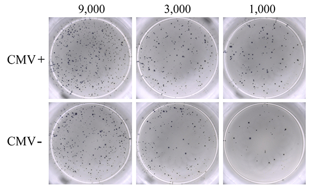
Figure 2.Representative images of filter plate wells show distinct spots that are counted with an ELISpot reader. These results are usually presented as bar graphs displaying spot-forming units (SFU) per million cells, providing single-cell resolution.
When to Choose ELISA
ELISA is ideal when your goal is to quantitate the concentration of a specific protein(s) in solution. Whether you're working with serum, plasma, cell culture supernatants, or other fluids, ELISA provides a reliable and well-established platform for detecting soluble analytes.
Select ELISA when:
- You need to determine protein concentration (pg/mL, ng/mL, etc.)
- The target protein is a secreted protein (cytokines, antibodies, or hormones)
- Your experiment involves comparative analysis across multiple samples, such as treatment groups or time points
- You need a high-throughput, scalable solution with automation compatibility
- You’re working with limited numbers of cells or simply don't need cellular resolution
Typical applications include:
- Profiling cytokine secretion in response to stimuli
- Measuring antibody titers in infection or vaccine studies
- Monitoring biomarkers in clinical samples
- Validating protein expression levels
When to Choose ELISpot
ELISpot is ideal when you need to evaluate functional immune response at the single-cell level. ELISpot is designed to detect and count individual cells that secrete target protein, offering unmatched sensitivity for identifying rare responding cells.
Choose ELISpot when:
- You want to measure the number of target protein-secreting cells (e.g., IFNγ-producing T cells)
- You need to assess cell-mediated immunity, particularly in response to antigens or pathogens
- You're working with PBMCs, isolated immune cells, or splenocytes from human or animal sources
- You require high sensitivity
Typical applications include:
- Monitoring T cell response in vaccine trials
- Evaluating immune activation in infectious disease or oncology studies
- Tracking B cell antibody secretion at the single-cell level
- Detecting rare immune events that are not discernible in bulk protein analysis
Practical Considerations
When deciding between ELISA and ELISpot, it's also important to consider some practical aspects of your experiment:
- Sample type: ELISA requires a liquid sample (e.g., serum, plasma, or cell culture supernatant). ELISpot requires freshly isolated or properly frozen immune cells, which may limit its use.
- Sensitivity: ELISpot offers single-cell resolution and can detect rare antigen-specific responses. ELISA is less sensitive but provides robust concentration profiles for proteins that are more abundantly secreted.
- Detection equipment: ELISA results are typically measured using standard plate readers. ELISpot requires a specialized plate reader and analysis software to detect and count individual spots (cells).
- Assay complexity: ELISA protocols tend to be more streamlined and simpler. ELISpot requires careful cell handling, making it slightly more technically demanding.
- Results: ELISA gives you a quantitative protein concentration (e.g., pg/mL), while ELISpot gives you the number of secreting cells (e.g., SFU per 10⁶ cells).
Using ELISA and ELISpot Together
While ELISA and ELISpot each offer unique strengths, they are highly complementary when used together. For example, ELISpot can tell you how many cells are producing a certain cytokine, whereas ELISA can tell you how much of that cytokine ends up in the supernatant. By pairing these assays, one can build a more complete picture of cellular response.
- Vaccine development: Use ELISpot to detect antigen-specific T cell responses, and ELISA to quantify antibody titers or secreted cytokines in serum.
- Immunotherapy monitoring: Use ELISpot to track functional immune activation at the cellular level, and ELISA to monitor systemic cytokine release.
- Biomarker discovery: Use ELISpot to identify rare immune responders, and ELISA to confirm downstream protein levels in fluid samples.
Researchers often find that combining ELISA and ELISpot can offer deeper insight into immune response.