Millipore Sigma Vibrant Logo
Attenzione: Ci siamo trasferiti. Non è più possibile acquistare i prodotti Merck Millipore nel sito MerckMillipore.comPer saperne di più


AXIS™ Axon Isolation Device

Request Information

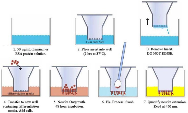
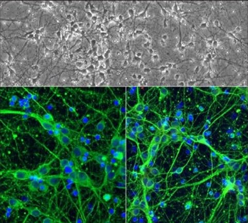
AXIS™ axon isolation device.The AXIS™ platform is Merck’s most advanced tool for the study of neurite outgrowth. This slide-mounted microfluidic chamber system enables the deposition and culture of neural cells and the spatially controlled addition of growth factors, toxins, and other reagents. Neurite outgrowth is restricted to narrow, parallel channels, and the resultant outgrowth or collapse behavior is easily observed under a microscope. The result is a powerful platform for the study of somas, neurites or synaptic formation.

The AXIS™ Axon Isolation Device is a two chamber system, each composed of two wells and an interconnected channel, separated by a set of microgrooves. The hydrostatic pressure formed by volume differential between chambers induces fluidic isolation of the solution on the low volume side of the device. The microfluidic design of an AXIS™ device allows for development and maintenance of a fluidic gradient of chemoattractants, toxins or other molecules of interest, facilitating controlled exposure and differentiation of axons.

Key Highlights

  • Organize, visualize, and characterize neuronal cell culture.
  • Detect protein expression with better spatial resolution.Isolate cell bodies from axons through fluidics.
  • Reduce time and expense through optimized protocols and QC validated products.
  • Attain superior performance over in-house protocols.
  • Optically clear transparent, inert, non-toxic, and nonflammable polymer mold.
  • Available in 150 μm, 450 μm, 900 μm, or 6-well.

Ordering Information


How the AXIS™ Isolation Device Works


Merck:/Freestyle/BI-Bioscience/Cell-Culture/new-cell-culture-images/VolumeMediated-HydrostaticFlow.jpg

Merck:/Freestyle/BI-Bioscience/Cell-Culture/cell-culture-images/triple-staining.bmp

Supporting Data

AXIS™: Axon Investigation System - Spatially Controlled Outgrowth Platform
Nervous system development results in a network of synaptic connections between participating neurons. Understanding the process of formation of this network is crucial to improving therapeutic treatments for patients suffering from nervous system developmental disorders and from neurodegenerative diseases, such as Parkinson’s disease, Huntington’s disease, and Lewy body disease1.

Neural connections are made when neuron filaments grow outwards in response to axon guidance cues, which include extracellular chemoattractant gradients and intracellular signaling molecules2.

Historically, neurite outgrowth assays have been able to detect only gross correlations between signaling events, molecular gradients and axon growth. From these studies, it has been shown that proteins such as F-actin and microtubules accumulate in the neuron’s growth cone in response to axon growth cues3.

Recent research has focused on studying the causes of directionality of axon growth. The polarity of axon growth coincides with gradients of extracellular signals, but it is not yet clear how extracellular gradients translate into asymmetric distribution and function of intracellular proteins driving neurite outgrowth4.

To fully understand the biochemical mechanisms by which axons grow in response to signaling, one should assay each axon, in isolation from somas and other neurons.

Spatial studies of directional neuronal outgrowth have been hampered by a lack of a solid, flexible, neurite culturing platform.

Traditionally, outgrowth experiments are conducted either by culturing neural stem cells (NSCs), primary neurons, or tissue slices with standard cultureware. This leads to random neurite outgrowth or outgrowth in particular directions guided by specific growth factors as dictated by the experimental design. Even in growth-factor-guided situations, neurite outgrowth is often crowded and somewhat haphazard. The ability to spatially isolate and study individual neurites is difficult in these preparations due to erratic growth, clumping and bifurcation. A simple, inexpensive, repeatable way to grow and isolate neurites would greatly enhance qualitative and quantitative studies.

The AXIS™ platform is Merck’s most advanced tool for the study of neurite outgrowth.

References:
  1. Yaron, A. and Zheng, B. (2007). Navigating their way to the clinic: emerging roles for axon guidance molecules in neurological disorders and injury. Dev. Neurobiol. 67:1216-1231.
  2. Chilton JK. Molecular mechanisms of axon guidance. Dev Biol. 2006 Apr 1; 292(1):13-24.
  3. Lin CH and Forscher P. Cytoskeletal remodeling during growth cone-target interactions. J Cell Biol. 1993 Jun;121(6):1369-1383.
  4. Quinn CC and Wadsworth WG. Axon guidance: asymmetric signaling orients polarized outgrowth. Trends Cell Biol. 2008 Oct; 18(12):597-603.