


OP80 Sigma-AldrichAnti-APC (Ab-7) Mouse mAb (CC-1)


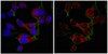
Anti-APC (Ab-7), mouse monoclonal, clone CC-1, recognizes APC in oligodendrocytes and astrocytes. It is validated for ICC, IF, free-floating Sections, and paraffin and frozen sections.
More>> Anti-APC (Ab-7), mouse monoclonal, clone CC-1, recognizes APC in oligodendrocytes and astrocytes. It is validated for ICC, IF, free-floating Sections, and paraffin and frozen sections. Less<<Anti-APC (Ab-7) Mouse mAb (CC-1) MSDS (material safety data sheet) or SDS, CoA and CoQ, dossiers, brochures and other available documents.
Synonyms: Anti-Adenomatus Polyposis Coli
Recommended Products


개요
| Replacement Information |
|---|
주요 사양표
| Species Reactivity | Host | Antibody Type |
|---|---|---|
| H, M, R | M | Monoclonal Antibody |
가격 및 재고여부
| 카탈로그 번호 | 재고 정보 | 패킹 | 포장 단위 | 가격(VAT 별도) | 수량 | |
|---|---|---|---|---|---|---|
| OP80-100UGCN |
|
Plastic ampoule | 100 μg |
|
— |
| Description | |
|---|---|
| Overview | Recognizes APC in oligodendrocytes and astrocytes.
|
| Catalogue Number | OP80 |
| Brand Family | Calbiochem® |
| Synonyms | Anti-Adenomatus Polyposis Coli |
| Product Information | |
|---|---|
| Form | Liquid |
| Formulation | In 50 mM sodium phosphate buffer, 0.2% gelatin. |
| Negative control | MOPC 21 cells |
| Positive control | Purkinje cells or cerebellum tissue |
| Preservative | ≤0.1% sodium azide |
| Quality Level | MQ100 |
| Physicochemical Information |
|---|
| Dimensions |
|---|
| Materials Information |
|---|
| Toxicological Information |
|---|
| Safety Information according to GHS |
|---|
| Safety Information |
|---|
| Product Usage Statements |
|---|
| Storage and Shipping Information | |
|---|---|
| Ship Code | Blue Ice Only |
| Toxicity | Standard Handling |
| Storage | +2°C to +8°C |
| Do not freeze | Yes |
| Packaging Information |
|---|
| Transport Information |
|---|
| Supplemental Information |
|---|
| Specifications |
|---|
| Global Trade Item Number | |
|---|---|
| 카탈로그 번호 | GTIN |
| OP80-100UGCN | 04055977209662 |
Documentation
Anti-APC (Ab-7) Mouse mAb (CC-1) Certificates of Analysis
| Title | Lot Number |
|---|---|
| OP80 |
References
| 참고문헌 보기 |
|---|
| Bhat, R.V., et al. 1996. Glia 17, 169. Bhat, R.V., et al. 1994. Neuroscience Proceedings. Smith, K.J., et al. 1994. Cancer Res. 54, 3672. Smith, K.J., et al. 1993. Proc. Natl. Acad. Sci. USA 90, 2846. Su, L.-K., et al. 1993. Cancer Res. 53, 2728. Su, L.-K., et al. 1993. Science 262, 1734. Groden, J., et al. 1991. Cell 66, 589. Kinzler, K.W., et al. 1991. Science 251, 1366. |