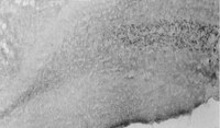
BACE1-/- mice exhibit seizure activity that does not correlate with sodium channel level or axonal localization.
Hitt, BD; Jaramillo, TC; Chetkovich, DM; Vassar, R
Molecular neurodegeneration
5
31
2010
Show Abstract
BACE1 is a key enzyme in the generation of the Abeta peptide that plays a central role in the pathogenesis of Alzheimer's disease. While BACE1 is an attractive therapeutic target, its normal physiological function remains largely unknown. Examination of BACE1-/- mice can provide insight into this function and also help anticipate consequences of BACE1 inhibition. Here we report a seizure-susceptibility phenotype that we have identified and characterized in BACE1-/- mice.We find that electroencephalographic recordings reveal epileptiform abnormalities in some BACE1-/- mice, occasionally including generalized tonic-clonic and absence seizures. In addition, we find that kainic acid injection induces seizures of greater severity in BACE1-/- mice relative to BACE1+/+ littermates, and causes excitotoxic cell death in a subset of BACE1-/- mice. This hyperexcitability phenotype is variable and appears to be manifest in approximately 30% of BACE1-/- mice. Finally, examination of the expression and localization of the voltage-gated sodium channel alpha-subunit Nav1.2 reveals no correlation with BACE1 genotype or any measure of seizure susceptibility.Our data indicate that BACE1 deficiency predisposes mice to spontaneous and pharmacologically-induced seizure activity. This finding has implications for the development of safe therapeutic strategies for reducing Abeta levels in Alzheimer's disease. Further, we demonstrate that altered sodium channel expression and axonal localization are insufficient to account for the observed effect, warranting investigation of alternative mechanisms. | 20731874
 |
Voltage-gated sodium channels in taste bud cells.
Gao, N; Lu, M; Echeverri, F; Laita, B; Kalabat, D; Williams, ME; Hevezi, P; Zlotnik, A; Moyer, BD
BMC neuroscience
10
20
2009
Show Abstract
Taste bud cells transmit information regarding the contents of food from taste receptors embedded in apical microvilli to gustatory nerve fibers innervating basolateral membranes. In particular, taste cells depolarize, activate voltage-gated sodium channels, and fire action potentials in response to tastants. Initial cell depolarization is attributable to sodium influx through TRPM5 in sweet, bitter, and umami cells and an undetermined cation influx through an ion channel in sour cells expressing PKD2L1, a candidate sour taste receptor. The molecular identity of the voltage-gated sodium channels that sense depolarizing signals and subsequently initiate action potentials coding taste information to gustatory nerve fibers is unknown.We describe the molecular and histological expression profiles of cation channels involved in electrical signal transmission from apical to basolateral membrane domains. TRPM5 was positioned immediately beneath tight junctions to receive calcium signals originating from sweet, bitter, and umami receptor activation, while PKD2L1 was positioned at the taste pore. Using mouse taste bud and lingual epithelial cells collected by laser capture microdissection, SCN2A, SCN3A, and SCN9A voltage-gated sodium channel transcripts were expressed in taste tissue. SCN2A, SCN3A, and SCN9A were expressed beneath tight junctions in subsets of taste cells. SCN3A and SCN9A were expressed in TRPM5 cells, while SCN2A was expressed in TRPM5 and PKD2L1 cells. HCN4, a gene previously implicated in sour taste, was expressed in PKD2L1 cells and localized to cell processes beneath the taste pore.SCN2A, SCN3A and SCN9A voltage-gated sodium channels are positioned to sense initial depolarizing signals stemming from taste receptor activation and initiate taste cell action potentials. SCN2A, SCN3A and SCN9A gene products likely account for the tetrodotoxin-sensitive sodium currents in taste receptor cells. | 19284629
 |
Traumatic axonal injury induces proteolytic cleavage of the voltage-gated sodium channels modulated by tetrodotoxin and protease inhibitors.
Iwata, A; Stys, PK; Wolf, JA; Chen, XH; Taylor, AG; Meaney, DF; Smith, DH
The Journal of neuroscience : the official journal of the Society for Neuroscience
24
4605-13
2004
Show Abstract
We demonstrated previously that dynamic stretch injury of cultured axons induces structural changes and Ca2+ influx modulated by tetrodotoxin (TTX)-sensitive voltage-gated sodium channels (NaChs). In the present study, we evaluated potential damage to the NaCh alpha-subunit, which can cause noninactivation of NaChs. In addition, we explored the effects of pre-injury and post-injury treatment with TTX and protease inhibition on proteolysis of the NaCh alpha-subunit and intra-axonal calcium levels ([Ca2+]i) over 60 min after trauma. After stretch injury, we found that [Ca2+]i continued to increase in untreated axons for at least 60 min. We also observed that the III-IV intra-axonal loop of the NaCh alpha-subunit was proteolyzed between 5 and 20 min after trauma. Pre-injury treatment of the axons with TTX completely abolished the posttraumatic increase in [Ca2+]i and proteolysis of the NaCh alpha-subunit. In addition, both pre-injury and post-injury inhibition of protease activity attenuated long-term increases in [Ca2+]i as well as mitigating degradation of the NaCh alpha-subunit. These results suggest a unique "feed-forward" deleterious process initiated by mechanical trauma of axons. Na+ influx through NaChs resulting from axonal deformation triggers initial increases in [Ca2+]i and subsequent proteolysis of the NaCh-subunit. In turn, degradation of the alpha-subunit promotes persistent elevations in [Ca2+]i, fueling additional pathologic changes. These observations may have important implications for developing therapeutic strategies for axonal trauma. | 15140932
 |
Elevated expression of type II Na+ channels in hypomyelinated axons of shiverer mouse brain.
Westenbroek, R E, et al.
J. Neurosci., 12: 2259-67 (1992)
1992
Show Abstract
Type I and type III Na+ channels are localized mainly in neuronal cell bodies in mouse brain. Type II channels are preferentially localized in unmyelinated fiber tracts but are not detectable in normally myelinated fibers. In shiverer mice, which lack compact myelin due to a defect in the myelin basic protein gene, elevated expression of type II Na+ channels was observed in the hypomyelinated axons of large-caliber fiber tracts such as the corpus callosum, internal capsule, fimbria, fornix, corpus medullare of the cerebellum, and nigrostriatal pathway by immunocytochemical analysis with subtype-specific antibodies. No difference was observed in the localization of type I and type III Na+ channels between wild-type and shiverer mice. These findings support the hypothesis that type II Na+ channels are preferentially localized in axons of brain neurons and suggest that their density and localization are regulated by myelination. The selective increase in the number of type II channels in hypomyelinated fiber tracts may contribute to the hyperexcitable phenotype of the shiverer mouse. | 1318958
 |
Tissue-specific expression of the RI and RII sodium channel subtypes.
Gordon, D, et al.
Proc. Natl. Acad. Sci. U.S.A., 84: 8682-6 (1987)
1987
Show Abstract
Anti-peptide antibodies that distinguish between the rat brain sodium channel subtypes referred to as RI and RII were prepared and used to determine their relative expression in nerve and muscle tissues. Sodium channels purified from rat brain are approximately 18% RI and 80% RII. In brain, the RII subtype is preferentially expressed with RI/RII ratios ranging from 0.07 in the hippocampus to 0.17 in the cerebral cortex. The RI subtype is preferentially expressed in more caudal areas of the central nervous system with values of RI/RII of 0.98 for medulla oblongata and 2.2 for spinal cord. Expression of additional unidentified sodium channel subtype(s) is detected in midbrain, medulla, and spinal cord, and expression of unidentified sodium channel subtypes predominates over expression of RI and RII in retina and optic nerve. The RI and RII subtypes are primarily expressed in the central nervous system and are not detected in significant numbers in skeletal or cardiac muscle, sympathetic ganglia, adrenal medulla, sciatic nerve, or cauda equina. The RII subtype appears first in development of both brain and spinal cord but declines in adult spinal cord as the RI subtype increases. The strict regional expression of these two sodium channel subtypes suggests that they may have distinct functional properties or physiological roles. | 2446328
 |
Expression of functional sodium channels from cloned cDNA.
Noda, M, et al.
Nature, 322: 826-8 (1986)
1986
Show Abstract
The voltage-gated sodium channel is a transmembrane protein essential for the generation of action potentials in excitable cells. It has been reported that sodium channels purified from the electric organ of the electric eel, Electrophorus electricus, and from chick cardiac muscle consist of a single polypeptide of relative molecular mass (Mr) approximately 260,000, whereas those purified from rat brain and from rat and rabbit skeletal muscle contain, in addition to the large polypeptide, one or two smaller polypeptides of Mr 33,000-43,000. The primary structures of the Electrophorus sodium channel and two distinct sodium channel large polypeptides (designated as sodium channels I and II) from rat brain have been elucidated by cloning and sequencing the complementary DNAs. The purified sodium channel preparations from Electrophorus electroplax and from mammalian muscle and brain, when reconstituted into lipid vesicles or planar lipid bilayers, exhibit some functional activities. The successful reconstitution with the Electrophorus preparation would imply that the large polypeptide alone is sufficient to form functional sodium channels. However, studies with the rat brain preparation suggest that the smaller polypeptide of Mr 36,000 is also required for the integrity of the saxitoxin (STX) or tetrodotoxin (TTX) binding site of the sodium channel. Here we report that the messenger RNAs generated by transcription of the cloned cDNAs encoding the rat brain sodium channel large polypeptides, when injected into Xenopus oocytes, can direct the formation of functional sodium channels. | 2427955
 |


