
113000
Acridine Orange - CAS 65-61-2 - Calbiochem
Synonym(s):
Acridine Orange - CAS 65-61-2 - Calbiochem, AO, 3,6- bis(Dimethylamino)acridine, HCl
Sign In to View Organizational & Contract Pricing.
Select a Size
Change View
About This Item
CAS Number:
UNSPSC Code:
12352200
assay
≥98% (HPLC)
form
solid
manufacturer/tradename
Calbiochem®
storage condition
OK to freeze, desiccated (hygroscopic), protect from light
color
burnt orange
solubility
DMF: 1 mg/mL, H2O: 1 mg/mL, ethanol: 1 mg/mL
shipped in
ambient
storage temp.
15-25°C
InChI
1S/C17H19N3.ClH/c1-19(2)13-8-9-15-12(10-13)11-14-16(18-15)6-5-7-17(14)20(3)4;/h5-11H,1-4H3;1H
InChI key
ASKSWIKIIFUEOI-UHFFFAOYSA-N
General description
A cell-permeable fluorescent dye that interacts with DNA and RNA by intercalation or electrostatic attractions. When bound to DNA, this cationic dye is spectrally very similar to fluorescein, with an excitation maximum at 502 nm and an emission maximum at 525 nm. Upon association with RNA, the excitation maximum shifts to 460 nm and the emission maximum shifts to 650 nm (red). Often used to measure single- and double-stranded DNA and RNA in the diagnosis, classification, and prognostication of many neoplasms. A very versatile fluorescent stain used in histochemistry and cytochemistry providing information about the in situ content, molecular structure, conformation, and environment of many nucleic acid-containing cell constituents.
A cell-permeable, cationic fluorescent dye that interacts with DNA and RNA by intercalation or electrostatic attractions. When bound to DNA, it is spectrally very similar to fluorescein, with an excitation maximum at 502 nm and an emission maximum at 525 nm. Upon association with RNA, the excitation maximum shifts to 460 nm and the emission maximum shifts to 650 nm (red). Often used to measure single- and double-stranded DNA and RNA in the diagnosis, classification, and prognosis of many neoplasms. A very versatile fluorescent stain used in histochemistry and cytochemistry that can be used to provide information about the in situ content, molecular structure, and conformation of many nucleic acid-containing cell constituents.
Biochem/physiol Actions
Cell permeable: yes
Primary Target
Interacts with DNA and RNA
Interacts with DNA and RNA
Product does not compete with ATP.
Preparation Note
Following reconstitution, store in the refrigerator (4°C). Aqueous stock solutions are stable for up to 1 month at 4°C.
Other Notes
Gonzalez, K., et al. 1995. Curr. Eye Res.14, 269.
Muro-Cacho, C.A., et al. 1995. J. Immunol.154, 5555.
Olivier, R. 1995. Methods Enzymol.251, 270.
Busch, G.L., et al. 1994. Proc. Natl. Acad. Sci. USA91, 9165.
Darzynkiewicz, Z. 1994. Methods Cell. Biol.41, 401.
Delic, J., et al. 1991. Exp. Cell Res.194, 147.
El-Naggar, A.K., et al. 1991. Cytometry12, 330.
Lopez, F., et al. 1991. Cytometry12, 42.
Gurrieri, S., et al. 1990. Biochemistry29, 3396.
Hermansen, D.K., et al. 1989. Cytometry10, 739.
Legal Information
CALBIOCHEM is a registered trademark of Merck KGaA, Darmstadt, Germany
Disclaimer
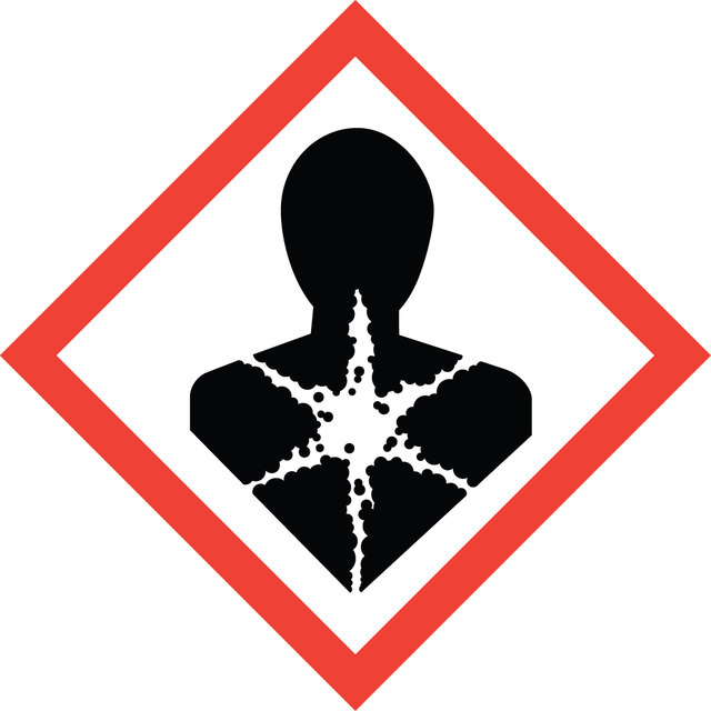
Toxicity: Harmful & Carcinogenic / Teratogenic (E)
signalword
Warning
hcodes
Hazard Classifications
Muta. 2
Storage Class
11 - Combustible Solids
wgk
WGK 3
flash_point_f
Not applicable
flash_point_c
Not applicable
Certificates of Analysis (COA)
Search for Certificates of Analysis (COA) by entering the products Lot/Batch Number. Lot and Batch Numbers can be found on a product’s label following the words ‘Lot’ or ‘Batch’.
Already Own This Product?
Find documentation for the products that you have recently purchased in the Document Library.