콘텐츠로 건너뛰기
Merck
HomeCell Counting & Health Analysis세포 생존성 및 증식을 위한 MTT 분석 프로토콜

세포 생존성 및 증식을 위한 MTT 분석 프로토콜

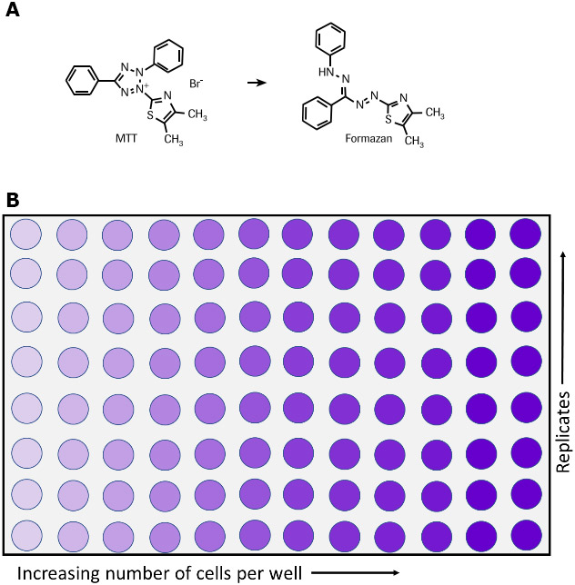
MTT 분석은 세포 생존성, 증식, 세포독성의 지표로서 세포 대사 활성을 측정하는 데 사용됩니다. 이 비색분석은 노란색 tetrazolium 염(3-(4,5-dimethylthiazol-2-yl)-2,5-diphenyltetrazolium bromide 또는 MTT)이 대사적으로 활성화된 세포에 의해 자주색 formazan 결정으로 환원되는 것에 기반합니다(그림 1).6,7,35 생육 가능한 세포에는 MTT를 formazan으로 환원시키는 NAD(P)H 의존성 산화환원효소가 함유되어 있습니다.36 불용성 formazan 결정은 용해화 용액을 사용하여 용해시킬 수 있으며, 그 결과로 생성된 색상이 있는 용액은 다중 웰 분광광도계를 사용하여 500~600 nm에서 흡광도 측정을 통해 정량화됩니다. 용액의 색상이 어두울수록 생육 가능하고 대사적으로 활성인 세포의 숫자가 더 많아집니다.

MTT를 사용하는 이 비방사성 비색분석 시스템은 Mosmann, T 외1에 의해 처음 기술되었으며 이후 몇 년 동안 여러 다른 연구자에 의해 개선되었습니다.2-6 Cell Proliferation Kit I (MTT)는 즉시 사용 가능한 시약을 포함한 최적화된 MTT 분석 키트이며, 수세 단계나 추가적인 시약이 필요하지 않습니다. 많은 수의 시료를 신속하고 편리하게 처리할 수 있는 정량 분석입니다. Cell Proliferation Kit I (MTT)는 다음과 같은 여러 응용분야에서 사용될 수 있습니다.

  • 세포 성장 및 생존성의 정량화.1,3,5-7
  • 성장 인자, 사이토카인, 영양소에 대한 반응으로 일어나는 세포 증식의 측정.1-3,6,8-12(그림 3 참조).
  • 세포독성 측정. 관련 예시로는 종양괴사인자-a 또는 -b 효과13,14(그림 2 참조) 또는 대식세포 유도 세포사15,16의 정량화, 그리고 세포독성 17-34 또는 성장 억제제(예: 억제 항체)에 대한 평가가 있습니다.
  • 세포 활성 연구.4
Formazan 염 및 96웰 플레이트에 대한 MTT의 대사

그림 1.화학 반응(A) 및 96웰 플레이트(B)에서 나타난 것처럼 생육 가능한 세포에 의한 formazan 염에 대한 MTT의 대사.

Cell Proliferation Kit I(MTT) 구성요소(제품 11465007001번)

MTT 시약

  • 즉시 사용 가능, 비멸균
  • MTT(3-[4,5-dimethylthiazol-2-yl]-2,5-diphenyltetrazolium bromide) (1X) 5 ml가 인산염완충식염수(PBS, 806552)에 5 mg/mL 농도로 들어있는 바이알 5개

용해화 용액

  • 1X, 즉시 사용 가능
  • 90 mL 3병

세포독성 측정에 대한 분석 프로토콜

추가적으로 필요한 시약:

  • 배양 배지. 예를 들면 10% 열 불활성화 FCS(소태아 혈청, 12106C), 2 mM 글루타민(G6392), 1 μg/mL 액티노마이신 C1(액티노마이신 D, A9415)이 들어있는 RPMI 1640(R0883).
  • 항생제가 사용되는 경우, 추가적으로 배지에 페니실린/스트렙토마이신 또는 겐타마이신을 추가합니다.
  • 종양괴사인자-α, 인간(hTNF-α)(10 μg/mL), 멸균(T6674).

프로토콜:

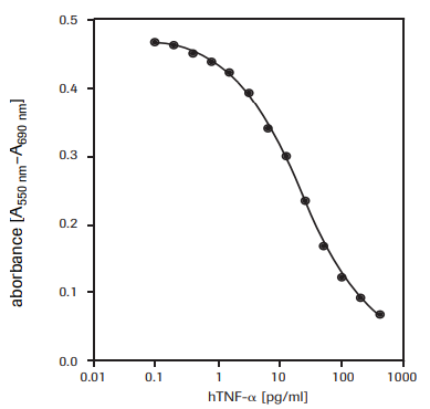
WEHI-164 세포(마우스 섬유육종, 87022501)에 대한 인간 종양괴사인자-α(hTNF-α, T6674)의 세포독성 효과를 측정하기 위함(그림 2 참조).

  1. 1 μg/mL 액티노마이신 C1이 포함된 배양 배지에서 1 × 10⁶ cells/ml 농도인 WEHI-164 세포를 37°C 및 5-6.5% CO2 조건 하에서 3시간 동안 사전배양합니다.
  2. 1 μg/ml 액티노마이신 C1 및 다양한 양의 hTNF-α(최종 농도 예: 0.001–0.5 ng/mL)가 들어있는 100 μl 배양 배지에서 농도가 5 × 10⁴ cells/ well인 세포를 마이크로플레이트(조직배양 등급, 96웰, 편평한 바닥) 안에 파종합니다.
  3. 37°C 이상 및 5-6.5% CO2 조건 하에서 24시간 동안 세포배양물을 배양합니다.
  4. 배양기간이 끝나면 각 웰에 MTT 라벨링 시약(최종 농도 0.5 mg/ml) 10 μl를 추가합니다.
  5. 습기가 있는 환경(예: 37°C 이상, 5-6.5% CO2)에서 마이크로플레이트를 4시간 동안 배양합니다.
  6. 각 웰에 용해화 용액 100 μl를 추가합니다.
  7. 습기가 있는 환경(예: 37°C 이상, 5-6.5% CO2)에 있는 배양기에 플레이트를 하룻밤 동안 둡니다.
  8. 자주색 formazan 결정의 완전한 용해화를 확인하고 마이크로플레이트(ELISA) 판독기를 사용하여 시료의 흡광도를 측정합니다. Formazan 생성물의 흡광도를 측정하는 파장은 ELISA 판독기에 유효한 사용된 필터에 따라 550-600 nm입니다. 참조 파장은 650 nm 이상이어야 합니다.
세포독성 활성도의 측정

그림 2.WEHI-164 세포(마우스 섬유육종)에 대한 재조합형 인간 TNF-α(hTNF-α)의 세포독성 활성도를 MTT 분석으로 측정.

세포 성장 측정에 대한 분석 프로토콜

추가적으로 필요한 시약

  • 10% 열 불활성화 FCS(소태아 혈청, 제품번호 12106C)가 들어있는 배양 배지(예: DMEM, 제품번호 D5671)
  • 2 mM 글루타민(제품번호 G6392)
  • 0.55 mM L-asparagine(제품번호 A8094
  • 0.24 mM L-asparagine-monohydrate(제품번호 A4284)
  • 50 μM 2-mercaptoethanol(제품번호 M3148), 
  • HT 배지 보충제(제품번호 H0137) (1×), 0.1 mM hypoxanthine 및 16 μM thymidine 함유. 항생제가 사용되는 경우, 추가적으로 배지에 페니실린/스트렙토마이신 또는 겐타마이신을 추가합니다. 
  • 인터루킨-6, 인간(hIL-6, 제품번호 SRP3096)(200,000 U/mL, 2 μg/mL) 멸균

프로토콜

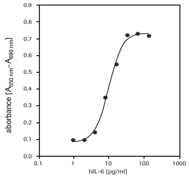
7TD1 세포(마우스-마우스 하이브리도마, DSMZ, ACC 23)에 대한 인간 인터루킨-6(hIL-6) 활성도 측정을 위함(그림 3 참고).

  1. 다양한 양의 IL-6[최종 농도 예: 0.1-10 U/mL(0.001-0.1 ng/mL)]가 들어있는 100 μL 배양 배지에서 농도가 2 × 10³ cells/well인 7TD1 세포를 마이크로플레이트(조직배양 등급, 96웰, 편평한 바닥) 안에 파종합니다.
  2. 37°C 이상 및 5-6.5% CO2 조건 하에서 4일 동안 세포배양물을 배양합니다.
  3. 배양기간이 끝나면 각 웰에 MTT 라벨링 시약(최종 농도 0.5 mg/mL) 10 μL를 추가합니다.
  4. 습기가 있는 환경(예: 37°C 이상, 5-6.5% CO2)에서 마이크로플레이트를 4시간 동안 배양합니다.
  5. 각 웰에 용해화 용액 100 μL를 추가합니다.
  6. 습기가 있는 환경(예: 37°C 이상, 5-6.5% CO2)에 있는 배양기에 플레이트를 하룻밤 동안 둡니다.
  7. 자주색 formazan 결정의 완전한 용해화를 확인하고 마이크로플레이트(ELISA) 판독기를 사용하여 분광광도법으로 시료의 흡광도를 측정합니다. Formazan 생성물의 흡광도를 측정하는 파장은 ELISA 판독기에 유효한 사용된 필터에 따라 550-600 nm입니다. 참조 파장은 650 nm 이상이어야 합니다.
7TD1 세포의 증식

그림 3.재조합형 인간 인터루킨-6(hIL-6)에 대한 반응으로서 7TD1 세포(마우스-마우스 하이브리도마)의 증식. MTT 분석 사용.

유사한 분석법

XTT 분석 및 WST-1 분석은 세포 생존성 및 증식을 측정할 때도 사용될 수 있습니다.

제품

Loading

참고문헌

1.
Mosmann T. 1983. Rapid colorimetric assay for cellular growth and survival: Application to proliferation and cytotoxicity assays. Journal of Immunological Methods. 65(1-2):55-63. https://doi.org/10.1016/0022-1759(83)90303-4
2.
Tada H, Shiho O, Kuroshima K, Koyama M, Tsukamoto K. 1986. An improved colorimetric assay for interleukin 2. Journal of Immunological Methods. 93(2):157-165. https://doi.org/10.1016/0022-1759(86)90183-3
3.
Denizot F, Lang R. 1986. Rapid colorimetric assay for cell growth and survival. Journal of Immunological Methods. 89(2):271-277. https://doi.org/10.1016/0022-1759(86)90368-6
4.
Gerlier D, Thomasset N. 1986. Use of MTT colorimetric assay to measure cell activation. Journal of Immunological Methods. 94(1-2):57-63. https://doi.org/10.1016/0022-1759(86)90215-2
5.
Hansen MB, Nielsen SE, Berg K. 1989. Re-examination and further development of a precise and rapid dye method for measuring cell growth/cell kill. Journal of Immunological Methods. 119(2):203-210. https://doi.org/10.1016/0022-1759(89)90397-9
6.
Vistica VT, Skehan P, Scudiero D, Monks A, Pittman A, Boyd MR. 1991. Tetrazolium-based assays for cellular viability: a critical examination of selected parameters affecting formazan production. Cancer Res. 51(10):2515-20.
7.
Maehara Y, Anai H, Tamada R, Sugimachi K. 1987. The ATP assay is more sensitive than the succinate dehydrogenase inhibition test for predicting cell viability. European Journal of Cancer and Clinical Oncology. 23(3):273-276. https://doi.org/10.1016/0277-5379(87)90070-8
8.
Heeg K, Reimann J, Kabelitz D, Hardt C, Wagner H. 1985. A rapid colorimetric assay for the determination of IL-2-producing helper T cell frequencies. Journal of Immunological Methods. 77(2):237-246. https://doi.org/10.1016/0022-1759(85)90036-5
9.
Hooton, Gibbs J, Paetkan U C. 1985. Interaction of interleukin 2 with cells: quantitative analysis of effects.. J Immunol. 1352464–2473.
10.
Mosmann T, Fong T. 1989. Specific assays for cytokine production by T cells. Journal of Immunological Methods. 116(2):151-158. https://doi.org/10.1016/0022-1759(89)90198-1
11.
Ohno M, Abe T. 1991. Rapid colorimetric assay for the quantification of leukemia inhibitory factor (LIF) and interleukin-6 (IL-6). Journal of Immunological Methods. 145(1-2):199-203. https://doi.org/10.1016/0022-1759(91)90327-c
12.
Berg K, Hansen MB, Nielsen SE. 1988. J. Interferon Res.. 8((suppl. 1)):S. 67.
13.
Green LM, Reade JL, Ware CF. 1984. Rapid colormetric assay for cell viability: Application to the quantitation of cytotoxic and growth inhibitory lymphokines. Journal of Immunological Methods. 70(2):257-268. https://doi.org/10.1016/0022-1759(84)90190-x
14.
Espevik T, Nissen-Meyer J. 1986. A highly sensitive cell line, WEHI 164 clone 13, for measuring cytotoxic factor/tumor necrosis factor from human monocytes. Journal of Immunological Methods. 95(1):99-105. https://doi.org/10.1016/0022-1759(86)90322-4
15.
Ferrari M, Fornasiero MC, Isetta AM. 1990. MTT colorimetric assay for testing macrophage cytotoxic activity in vitro. Journal of Immunological Methods. 131(2):165-172. https://doi.org/10.1016/0022-1759(90)90187-z
16.
van de Loosdrecht AA, Nennie E, Ossenkoppele GJ, Beelen RH, Langenhuijsen MM. 1991. Cell mediated cytotoxicity against U 937 cells by human monocytes and macrophages in a modified colorimetric MTT assay. Journal of Immunological Methods. 141(1):15-22. https://doi.org/10.1016/0022-1759(91)90205-t
17.
Cole S. 1986. Rapid chemosensitivity testing of human lung tumor cells using the MTT assay. Cancer Chemother. Pharmacol.. 17(3): https://doi.org/10.1007/bf00256695
18.
Carmichael J, DeGraff WG, Gazdar AF, Minna JD, Mitchell JB. 1987. Evaluation of a tetrazolium-based semiautomated colorimetric assay: assessment of chemosensitivity testing. Cancer Res. 47(4):936-42.
19.
Twentyman P, Luscombe M. 1987. A study of some variables in a tetrazolium dye (MTT) based assay for cell growth and chemosensitivity. Br J Cancer. 56(3):279-285. https://doi.org/10.1038/bjc.1987.190
20.
Park JG, Park BS, Steinberg SM, Carmichael J, Collins JM, Minna JD, Gazdar AF. 1987. Chemosensitivity testing of human colorectal carcinoma cell lines using a tetrazolium-based colorimetric assay. Cancer Res. 475875-9.

21.
1988. FFPS News. Oryx. 22(4):246-250. https://doi.org/10.1017/s0030605300022481
22.
Pieters R, Huismans D, Leyva A, Veerman A. 1988. Adaptation of the rapid automated tetrazolium dye based (MTT) assay for chemosensitivity testing in childhood leukemia. Cancer Letters. 41(3):323-332. https://doi.org/10.1016/0304-3835(88)90294-7
23.
McHaLe A, McHaLe L. 1988. Use of a tetrazolium based colorimetric assay in assessing photoradiation therapy in vitro. Cancer Letters. 41(3):315-321. https://doi.org/10.1016/0304-3835(88)90293-5
24.
Edmondson JM, Armstrong LS, Martinez AO. 1988. A rapid and simple MTT-based spectrophotometric assay for determining drug sensitivity in monolayer cultures. Journal of Tissue Culture Methods. 11(1):15-17. https://doi.org/10.1007/bf01404408
25.
Faircloth GT, Stewart D, Clement JJ. 1988. A simple screening procedure for the quantitative measurement of cytotoxicity to resting primary lymphocyte cultures. Journal of Tissue Culture Methods. 11(4):201-205. https://doi.org/10.1007/bf01407314
26.
Campling BG, Pym J, Galbraith PR, Cole SP. 1988. Use of the mtt assay for rapid determination of chemosensitivity of human leukemic blast cells. Leukemia Research. 12(10):823-831. https://doi.org/10.1016/0145-2126(88)90036-7
27.
Pieters R, Huismans D, Leyva A, Veerman A. 1989. Comparison of the rapid automated MTT-assay with a dye exclusion assay for chemosensitivity testing in childhood leukaemia. Br J Cancer. 59(2):217-220. https://doi.org/10.1038/bjc.1989.44
28.
Twentyman PR, Fox NE, Rees JKH. 1989. Chemosensitivity testing of fresh leukaemia cells using the MTT colorimetric assay. Br J Haematol. 71(1):19-24. https://doi.org/10.1111/j.1365-2141.1989.tb06268.x
29.
Plumb JA, Milroy R, Kaye SB. 1989. Effects of the pH dependence of 3-(4,5-dimethylthiazol-2-yl)-2,5-diphenyl-tetrazolium bromide-formazan absorption on chemosensitivity determined by a novel tetrazolium-based assay. Cancer Res. 494435–4440.
30.
Alley MC, Scudiero DA, Monks A, Hursey ML, Czerwinski MJ, Fine DL, Abbott BJ, Mayo JG, Shoemaker RH, Boyd MR. 1988. Feasibility of drug screening with panels of human tumor cell lines using a microculture tetrazolium assay. Cancer Res. 48589–601.
31.
Heo DS, Park JG, Hata K, Day R, Herberman RB, Whiteside TL. 1990. Evaluation of tetrazolium-based semiautomatic colorimetric assay for measurement of human antitumor cytotoxicity. Cancer Res. 503681–3690.
32.
Ruben RL, Neubauer RH. 1987. Semiautomated colorimetric assay for in vitro screening of anticancer compounds. Cancer Treat Rep. 711141–1149..
33.
Rubinstein LV, Shoemaker RH, Paull KD, Simon RM, Tosini S, Skehan P, Scudiero DA, Monks A, Boyd MR. 1990. Comparison of In Vitro Anticancer-Drug-Screening Data Generated With a Tetrazolium Assay Versus a Protein Assay Against a Diverse Panel of Human Tumor Cell Lines. JNCI Journal of the National Cancer Institute. 82(13):1113-1117. https://doi.org/10.1093/jnci/82.13.1113
34.
1991. Announcement. Utilitas. 3(2):330-330. https://doi.org/10.1017/s0953820800001254
35.
Slater T, Sawyer B, Sträuli U. 1963. Studies on succinate-tetrazolium reductase systems. Biochimica et Biophysica Acta. 77383-393. https://doi.org/10.1016/0006-3002(63)90513-4
36.
Berridge M, Tan A. 1993. Characterization of the Cellular Reduction of 3-(4,5-dimethylthiazol-2-yl)-2,5-diphenyltetrazolium bromide (MTT): Subcellular Localization, Substrate Dependence, and Involvement of Mitochondrial Electron Transport in MTT Reduction. Archives of Biochemistry and Biophysics. 303(2):474-482. https://doi.org/10.1006/abbi.1993.1311