Patient-derived organoids as an epithelial model for preclinical IBD research and drug development
The study of the intestinal epithelial barrier has been hampered by the lack of suitable in vitro model systems. Patient-derived organoids (PDOs) recapitulate cellular diversity and patient heterogeneity, providing a more relevant platform for studying the intestinal epithelium and its response to microbial or inflammatory challenges.
Here we present the characterization of inflammatory bowel disease (IBD) organoid biobank derived from Crohn’s disease and ulcerative colitis patients and its application for IBD drug development efforts.
Interested in patient-derived organoids for IBD? Contact an expert now!
Section overview
Inflammatory bowel disease PDO biobank overview
The HUB Organoids® IBD Biobank includes:
- 11 growth validated organoid models from Chron’s disease and ulcerative colitis patients
- Small intestine and colon (ileum, proximal colon, distal colon, rectum) organoids
- Organoid models from un-inflamed and inflamed regions
- PDOs from normal tissues available as controls
Our intestinal organoids represent stem cells and fully differentiated epithelial cell types.
The PDOs in the IBD biobank mimic in vivo differentiation when cultured in specialized organoid media.
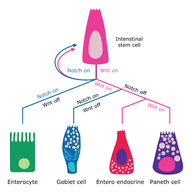
Figure 1.Cartoon representation of IBD differentiation.
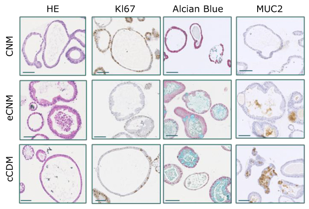
Figure 2.Microscopy images of the different cell types with different stains.
PDO monolayers can establish and differentiate on Corning® Transwell® inserts when cultured in specialized organoid media.
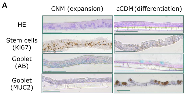
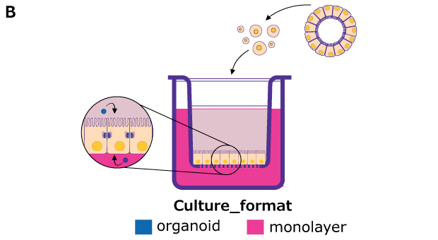
Figure 3. A) Immunohistochemistry images of the cell types stained for Ki67, AB, and MUC2 during expansion and differentiation; B) Cartoon representation of the culture format.
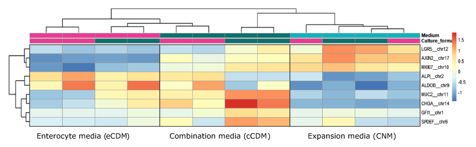
Figure 4.A heat map of protein expression in different cell types expressed in different media.
The intestinal cell types were enriched in different medium compositions (IHC, IF, and RNAseq data, see above).
The PDO intestinal monolayers are enriched for different cell types and allow for researchers to perform barrier studies and access the apical site.
Barrier integrity: cytokine-induced damage assay
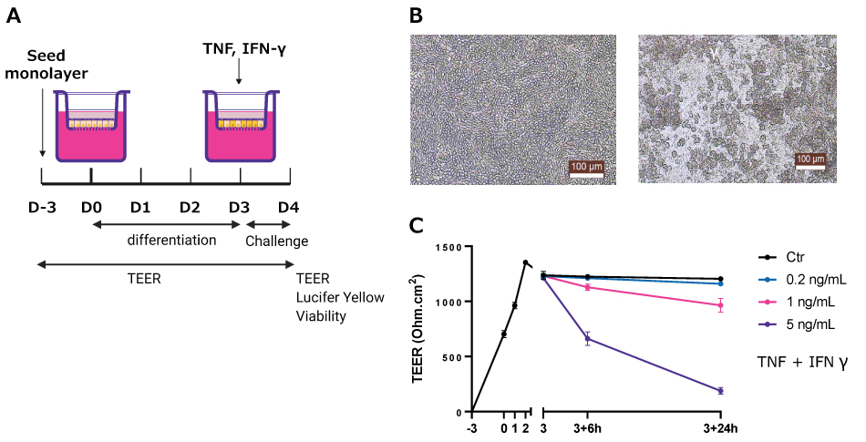
Figure 5.A) Cartoon representation of the experimental timeline; B) Microscopy images of the control and cytokine exposed monolayers; C) Graph of the TEER data from the monolayers.
The efficacy of Tofacitinib, an IBD therapy, on barrier protection is recapitulated in cytokine-induced damage assay.
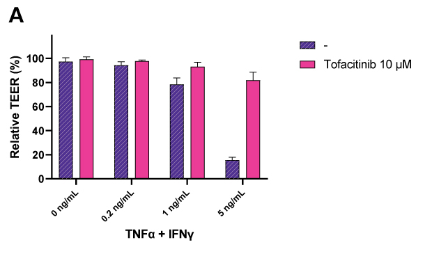
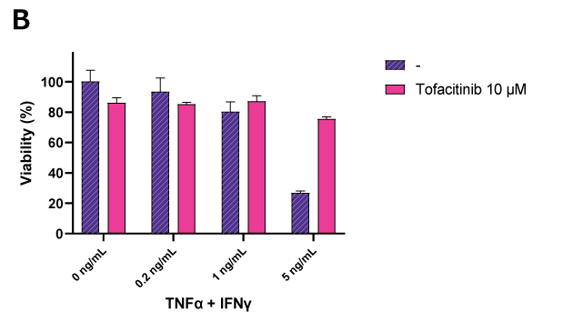
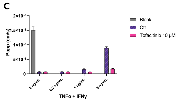
Figure 6. Graphs of barrier integrity data.A) barrier integrity, B) viability, and C) permeability cells exposed to Tofacitinib.
There is patient-to-patient variation in response to cytokine challenge, but there is a loss in barrier integrity across PDOs.
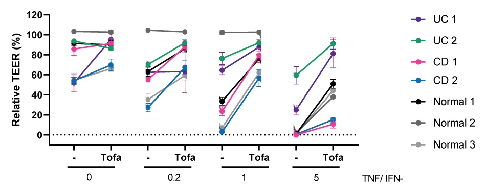
Figure 7.Graph showing the relative TEER percentage for cells exposed to Tofacitinib and different concentrations of TNF/IFNу.
Inflammatory bowel disease PDO biobank summary
PDOs recapitulate the cellular diversity and patient heterogeneity and provide a relevant platform to study the intestinal epithelium and its response to microbial or inflammatory challenges.
- Our IBD Biobank contains ulcerative colitis and Crohn’s disease patient samples.
- Specialized organoid media support the enrichment of distinct epithelial cell lineages.
- Intestinal PDO monolayers are enriched for differentiated cell types and allow barrier studies.
- Barrier damage during inflammatory conditions such as IBD can be studied with the cytokine-induced barrier damage assay.
- The cytokine challenge robustly induces loss of barrier integrity across seven patient-derived monolayers.
- The barrier breakdown was ameliorated in all models by treatment with Tofacitinib demonstrating the suitability of the assay for IBD drug development efforts.
For more information about our IBD biobank, IntergriGut PDO monolayer screen, or the IBD-diarrheic platform, check out our patient-derived organoids for inflammatory diseases page.