Millipore Sigma Vibrant Logo

CA1004 Anti-CA 125 Mouse mAb (OC 125)

View Products on Sigmaaldrich.com
CA1004
  
Retrieving price...
Price could not be retrieved
Minimum Quantity is a multiple of
Maximum Quantity is
Upon Order Completion More Information
You Saved ()
 
Request Pricing
Limited Availability
In Stock 
Discontinued
Limited Quantities Available
Availability to be confirmed
    Remaining : Will advise
      Remaining : Will advise
      Will advise
      Contact Customer Service
      Contact Customer Service


      Contact Customer Service

      Overview

      Replacement Information

      Key Spec Table

      Species ReactivityHostAntibody Type
      HMMonoclonal Antibody
      Description
      Overview

      This product has been discontinued.


      Recognizes the CA125 antigen on the >200 kDa mucin-like glycoprotein in non-mucinous epithelial ovarian carcinoma or uterine myoma.

      Catalogue NumberCA1004
      Brand Family Calbiochem®
      SynonymsAnti-Cancer Antigen 125
      Application Data
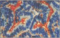
      Detection of human CA 125 by staining paraffin sections. Sample: Ovarian carcinoma fixed in formalin. Primary antibody: Anti-CA 125 Mouse mAb (OC 125) (Cat. No. CA1004) (1:40). Detection: DAB.
      References
      ReferencesHogdall, E.V., et al. 2002. APMIS 110, 545.
      Deng, X., et al. 2000. Gynecol. Oncol. 79, 416.
      Jeyarajah, A.R., et al. 1999. Gynecol. Oncol. 73, 242.
      Product Information
      FormLiquid
      FormulationIn 10 mM PBS, 1% BSA.
      Negative controlNormal mouse serum
      Positive controlNon-mucinous epithelial ovarian carcinoma, serous and endometroid adenocarcinomas, Krukenberg's tumor, or uterine myoma
      Preservative≤0.1% sodium azide
      Applications
      Application ReferencesFrozen Sections Fleuren, G.J., et al. 1987. Cancer 60, 2437. Argüeso, P., et al. 2003. Invest. Ophthamol. Vis. Sci. 44, 2487. Immunoblotting Lloyd, K.O., et al. 1997. Int. J. Cancer 71, 842. Selenmeyer, C., et al. 2003. J. Cell Sci. 116, 1305. Argüeso, P., et al. 2003. Invest. Ophthamol. Vis. Sci. 44, 2487. Immunoprecipitation Lloyd, K.O., et al. 1997. Int. J. Cancer 71, 842. Argüeso, P., et al. 2003. Invest. Ophthamol. Vis. Sci. 44, 2487. Immunocytochemistry Selenmeyer, C., et al. 2003. J. Cell Sci. 116, 1305.
      Key Applications Frozen Sections
      Immunoblotting (Western Blotting)
      Immunocytochemistry
      Immunoprecipitation
      Paraffin Sections
      Application NotesFrozen Sections (see application references)
      Immunoblotting (see application references)
      Immunoprecipitation (see application references)
      Immunocytochemistry (see application references)
      Paraffin Sections (1:40, heat pre-treatment required, see comments)
      Application CommentsWorks for formalin-fixed paraffin-embedded tissues. Pretreat with an antigen unmasking solution and heat for best results. Primary antibody may be diluted 1:40 for biotin based detection systems. For optimal staining, the primary antibody should be incubated 20-60 min at room temperature. Antibody should be titrated for optimal results in individual systems.
      Biological Information
      Immunogenovarian cancer cell line OVCA433
      ImmunogenHuman
      CloneOC 125
      HostMouse
      IsotypeIgG₁
      Species Reactivity
      • Human
      Antibody TypeMonoclonal Antibody
      Concentration Label Please refer to vial label for lot-specific concentration
      Physicochemical Information
      Dimensions
      Materials Information
      Toxicological Information
      Safety Information according to GHS
      Safety Information
      Product Usage Statements
      Storage and Shipping Information
      Ship Code Blue Ice Only
      Toxicity Highly Toxic
      Storage +2°C to +8°C
      Do not freeze Ok to freeze
      Special InstructionsFollowing initial use, aliquot and freeze (-20°C) for long-term storage if desired. Avoid freeze/thaw.
      Packaging Information
      Transport Information
      Supplemental Information
      Specifications
      Global Trade Item Number
      Catalogue Number GTIN
      CA1004 0

      Documentation

      Anti-CA 125 Mouse mAb (OC 125) Certificates of Analysis

      TitleLot Number
      CA1004

      References

      Reference overview
      Hogdall, E.V., et al. 2002. APMIS 110, 545.
      Deng, X., et al. 2000. Gynecol. Oncol. 79, 416.
      Jeyarajah, A.R., et al. 1999. Gynecol. Oncol. 73, 242.
      Data Sheet

      Note that this data sheet is not lot-specific and is representative of the current specifications for this product. Please consult the vial label and the certificate of analysis for information on specific lots. Also note that shipping conditions may differ from storage conditions.

      Revision09-July-2007 RFH
      SynonymsAnti-Cancer Antigen 125
      ApplicationFrozen Sections (see application references)
      Immunoblotting (see application references)
      Immunoprecipitation (see application references)
      Immunocytochemistry (see application references)
      Paraffin Sections (1:40, heat pre-treatment required, see comments)
      Application Data
      Detection of human CA 125 by staining paraffin sections. Sample: Ovarian carcinoma fixed in formalin. Primary antibody: Anti-CA 125 Mouse mAb (OC 125) (Cat. No. CA1004) (1:40). Detection: DAB.
      DescriptionPurified mouse monoclonal antibody. Recognizes the CA125 antigen on the >200 kDa mucin-like glycoprotein.
      BackgroundCancer antigen 125 is an antigenic determinant that is present on a high molecular weight, mucin-like glycoprotein of molecular weight greater than 200 kDa. It is common to most non-mucinous epithelial ovarian carcinomas. The CA 125 antigen is also present in other carcinomas. It is positive in serous and endometriod adenocarcinomas, Krukenberg's tumor, and uterine myoma. Mucinous tumors (benign, borderline, and malignant) and normal ovarian epithelium are negative. The CA 125 antigen is also detectable on the apical surface of normal endometrium and endocervix. Normal breast, skin, liver, spleen, and kidney are negative.
      HostMouse
      Immunogen speciesHuman
      Immunogenovarian cancer cell line OVCA433
      CloneOC 125
      IsotypeIgG₁
      Specieshuman
      Positive controlNon-mucinous epithelial ovarian carcinoma, serous and endometroid adenocarcinomas, Krukenberg's tumor, or uterine myoma
      Negative controlNormal mouse serum
      FormLiquid
      FormulationIn 10 mM PBS, 1% BSA.
      Concentration Label Please refer to vial label for lot-specific concentration
      Preservative≤0.1% sodium azide
      CommentsWorks for formalin-fixed paraffin-embedded tissues. Pretreat with an antigen unmasking solution and heat for best results. Primary antibody may be diluted 1:40 for biotin based detection systems. For optimal staining, the primary antibody should be incubated 20-60 min at room temperature. Antibody should be titrated for optimal results in individual systems.
      Storage +2°C to +8°C
      Do Not Freeze Ok to freeze
      Special InstructionsFollowing initial use, aliquot and freeze (-20°C) for long-term storage if desired. Avoid freeze/thaw.
      Toxicity Highly Toxic
      ReferencesHogdall, E.V., et al. 2002. APMIS 110, 545.
      Deng, X., et al. 2000. Gynecol. Oncol. 79, 416.
      Jeyarajah, A.R., et al. 1999. Gynecol. Oncol. 73, 242.
      Application referencesFrozen Sections Fleuren, G.J., et al. 1987. Cancer 60, 2437. Argüeso, P., et al. 2003. Invest. Ophthamol. Vis. Sci. 44, 2487. Immunoblotting Lloyd, K.O., et al. 1997. Int. J. Cancer 71, 842. Selenmeyer, C., et al. 2003. J. Cell Sci. 116, 1305. Argüeso, P., et al. 2003. Invest. Ophthamol. Vis. Sci. 44, 2487. Immunoprecipitation Lloyd, K.O., et al. 1997. Int. J. Cancer 71, 842. Argüeso, P., et al. 2003. Invest. Ophthamol. Vis. Sci. 44, 2487. Immunocytochemistry Selenmeyer, C., et al. 2003. J. Cell Sci. 116, 1305.