


17-10153 Sigma-AldrichLentiBrite™ RFP-Vimentin Lentiviral Biosensor


More>>
Less<<
Key Applications:
Transfection, Immunofluorescence, Immunocytochemistry, Track single cell and cell population migration (migration assay, cell culture)
MSDS (material safety data sheet) or SDS, CoA and CoQ, dossiers, brochures and other available documents.
Recommended Products
Overview
| Replacement Information |
|---|
Key Spec Table
| Key Applications | Detection Methods |
|---|---|
| TFX, IF, ICC, Track single cell and cell population migration (migration assay, cell culture) | Fluorescent |
| Description | |
|---|---|
| Catalogue Number | 17-10153 |
| Trade Name |
|
| Description | LentiBrite™ RFP-Vimentin Lentiviral Biosensor |
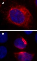
| Overview | Read our application note in Nature Methods! http://www.nature.com/app_notes/nmeth/2012/121007/pdf/an8620.pdf (Click Here!) Learn more about the advantages of our LentiBrite Lentiviral Biosensors! Click Here Biosensors can be used to detect the presence/absence of a particular protein as well as the subcellular location of that protein within the live state of a cell. Fluorescent tags are often desired as a means to visualize the protein of interest within a cell by either fluorescent microscopy or time-lapse video capture. Visualizing live cells without disruption allows researchers to observe cellular conditions in real time. Lentiviral vector systems are a popular research tool used to introduce gene products into cells. Lentiviral transfection has advantages over non-viral methods such as chemical-based transfection including higher-efficiency transfection of dividing and non-dividing cells, long-term stable expression of the transgene, and low immunogenicity. EMD Millipore is introducing LentiBrite™ Lentiviral Biosensors, a new suite of pre-packaged lentiviral particles encoding important and foundational proteins of autophagy, apoptosis, and cell structure for visualization under different cell/disease states in live cell and in vitro analysis.
EMD Millipore’s LentiBrite™ RFP-Vimentin lentiviral particles provide bright fluorescence and precise localization to enable live cell analysis of vimentin IF dynamics in difficult-to-transfect cell types. |
| Background Information | Intermediate filaments (IFs) are 10 nm diameter cytoskeletal structures that typically form a cage-like structure extending from the nucleus to the plasma membrane. The five families of IF proteins have distinct polymer characteristics and cell type-specific expression patterns. Expression of vimentin, a type-III IF protein, is restricted to mesenchymal cells, and appearance of vimentin in epithelial cancer cells is a hallmark of the epithelial-to-mesenchymal transition. Vimentin assembles into classic long filaments and short filaments termed squiggles. Time-lapse microscopy of live cells expressing RFP-vimentin has revealed the dynamics of vimentin IF maturation, and crosstalk with microtubules and microfilaments. EMD Millipore’s LentiBrite™ Vimentin-RFP lentiviral particles provide bright fluorescence and precise localization to enable live cell analysis of vimentin IF dynamics in difficult-to-transfect cell types. |
| References |
|---|
| Product Information | |
|---|---|
| Components |
|
| Detection method | Fluorescent |
| Quality Level | MQ100 |
| Biological Information | |
|---|---|
| Gene Symbol |
|
| Purification Method | PEG precipitation |
| UniProt Number | |
| Physicochemical Information |
|---|
| Dimensions |
|---|
| Materials Information |
|---|
| Toxicological Information |
|---|
| Safety Information according to GHS |
|---|
| Safety Information |
|---|
| Product Usage Statements | |
|---|---|
| Quality Assurance | Evaluated by transduction of HT-1080 cells and fluorescent imaging performed for assessment of transduction efficiency. |
| Usage Statement |
|
| Packaging Information | |
|---|---|
| Material Size | 1 vial (minimum of 3 x 10E8 IFU/mL) |
| Transport Information |
|---|
| Supplemental Information |
|---|
| Specifications |
|---|
| Global Trade Item Number | |
|---|---|
| Catalogue Number | GTIN |
| 17-10153 | 04053252633737 |
Documentation
LentiBrite™ RFP-Vimentin Lentiviral Biosensor SDS
| Title |
|---|
LentiBrite™ RFP-Vimentin Lentiviral Biosensor Certificates of Analysis
Technical Info
| Title |
|---|
| LentiBrite™ Lentiviral Biosensors for Fluorescent Cellular Imaging: Analysis of Autophagosome Formation |