
Immobilon® membranes for Western blotting
Western blotting requires extensive, multi-parameter optimization and antibody-based detection protocols that consume valuable sample and reagents. Immobilon® blotting membranes exhibit high protein binding capacity with superior retention across a broad range of protein sizes, including low-abundant proteins - minimizing time and conserving costly reagents while maximizing sensitivity and signal. We offer a wide selection of membrane chemistries and formats for your convenience.
View our Protein Blotting Handbook for more guidance.
Immobilon® transfer membranes for Western blotting
We offer five membranes:
- Immobilon®-NC Nitrocellulose membrane (0.45 μm) is an excellent substrate for immunodetection and is compatible with standard blocking agents and detection protocols.
- Immobilon®-P PVDF membrane (0.45 μm) is a versatile substrate that is well suited for common immunoblotting applications.
- Immobilon®-E PVDF membrane (0.45 μm) wets out in water or buffer, eliminating the alcohol pre-wet step while retaining similar performance to Immobilon®-P membranes.
- Immobilon®-PSQ PVDF membrane (0.2 μm) is ideal for protein sequencing and immunoblotting of low molecular weight proteins. It has a higher protein binding capacity and a higher retention than 0.45 μm membranes.
- Immobilon®-FL PVDF membrane (0.45 μm) was developed for fluorescence-based immunodetection. It has very low background fluorescence across a wide range of excitation and emission wavelengths.
Factors affecting protein binding in Western blotting
At the molecular level, protein adsorption results, at least in part, from the interaction of hydrophobic amino acid side chains and hydrophobic domains with the polymer surface. Matsudaira observed an 80% decline in the sequencing efficiency of small peptides after hydrophobic residues were cleaved, presumably due to the washout of the peptide remnants.1 Also, in peptide digestions, it has been observed that peptides characterized as hydrophobic often do not elute from the membrane as efficiently as more hydrophilic peptides.2, 3 McKeon and Lyman demonstrated that addition of Ca2+ ions to the transfer buffer enhanced the binding of calmodulin to the Immobilon®-P transfer membrane.4 Binding of the calcium to the protein causes formation of a hydrophobic pocket in the molecule’s structure, leading to enhanced binding to the hydrophobic PVDF surface.
Different membranes require different treatments for wetting out depending on whether the membrane is hydrophobic or hydrophilic. Traditional PVDF membranes will not wet out in aqueous solutions and must first be wet in ≥50%(v/v) solution of alcohol such as methanol, ethanol, or isopropanol. Immobilon®-E and Immobilon®-NC are hydrophilic and can be wet out in an aqueous solution such as transfer buffer or water. For all membranes, complete wetting is evident by a change in the membrane’s appearance from opaque to semi-transparent.
Binding differences between Immobilon®-P and Immobilon®-PSQ transfer membranes
After wetting, protein binding can be achieved by simply bringing the protein into contact with the membrane. Because binding occurs throughout the depth of the membrane, the binding capacity is determined by the internal surface area of the pores.5 Immobilon®-PSQ transfer membrane has approximately three times the internal surface area of Immobilon®-P transfer membrane, resulting in higher adsorptive capacity (Table 2). The values listed in Table 2 represent upper limits for protein binding after saturation of the membrane surface in a nondenaturing buffer. However, the maximum binding that can be achieved will depend on the specific protocols employed, variations in the structural conformation of the proteins, the chemical nature of the buffers used, and the limitations of the methods used to apply the sample. An example of the binding difference between Immobilon®-P and Immobilon®-PSQ transfer membranes is shown in Figure 2, where protein samples were electrotransferred from a polyacrylamide gel. A fraction of the proteins passed through the Immobilon®-P transfer membrane and were captured on a second membrane placed behind the first one. In contrast, all of the proteins were bound to the Immobilon®-PSQ membrane without passing through it. In this case, the tighter pore structure and higher internal membrane surface area facilitated complete adsorption of all of the transferred protein. However, immunodetection on Immobilon®-PSQ transfer membrane can result in a higher background and can require more stringent washing conditions.
Thus, membrane choice is dictated by the goal of the experiment; use Immobilon®-P transfer membrane for high-sensitivity detection of > 20 kDa proteins, but switch to Immobilon®-PSQ transfer membrane if smaller proteins are being analyzed or 100% protein capture is necessary for peptide sequencing.
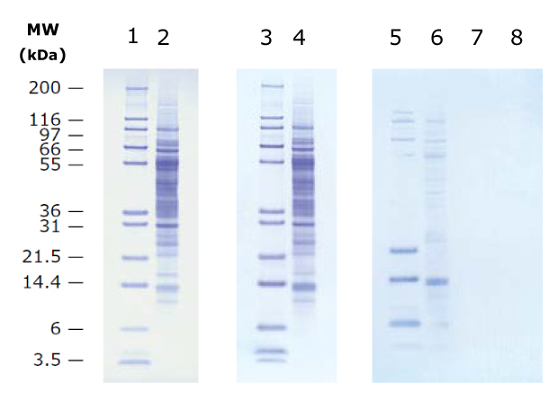
Figure 1.Prolonged electrotransfer of proteins using Immobilon®-P and Immobilon®-PSQ transfer membranes. Molecular weight standards (lanes 1,3,5,7) and calf liver lysate (lanes 2,4,6,8) were transferred to Immobilon®-P (lanes 1-2) or Immobilon®-PSQ (lanes 3-4) membranes by the tank transfer method and stained with Coomassie® Blue dye. A sheet of Immobilon®-PSQ transfer membrane was placed behind the primary membranes to capture proteins that passed through them (lanes 5 and 6 behind Immobilon®-P; lanes 7 and 8 behind Immobilon®-PSQ).
Immobilon® NOW membrane format
Immobilon® NOW is a pre-measured membrane roll format designed to streamline Western blot workflows. Available in five membrane types, Immobilon®-E, Immobilon®-P, Immobilo®-FL, Immobilon®-PSQ, and Immobilon®-NC, Immobilon® NOW rolls provide standardized mini and midi sizes with one dimension fixed at 8.5 cm, enabling direct substitution for cut sheets in applications where this dimension is applicable.
Standard transfer membrane rolls require manual measurement and cutting to achieve desired experimental sizes. This process introduces several practical considerations: extended bench time, potential material loss from measurement errors, and storage of partial rolls. Immobilon® NOW addresses these considerations by providing pre-measured formats that maintain membrane quality across standard blotting applications while reducing preparation time.
Immobilon® NOW rolls feature printed measurement marks on the container lid, allowing direct measurement without additional tools. The format is available in standard mini and midi sizes, with the 8.5 cm dimension permitting use as a direct replacement for pre-cut sheets in compatible protocols. The Immobilon® NOW Dispenser facilitates reproducible measurement and cutting of transfer membranes. The dispenser is recommended for use with Immobilon®-E, Immobilon®-P, and Immobilon®-PSQ membrane types. The integrated dispenser provides precise cutting capability, reduces handling of unused membrane through retraction storage, and maintains visibility of remaining material for inventory management.
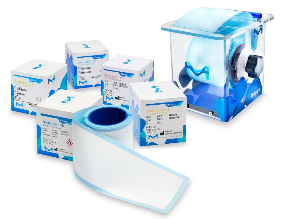
Figure 2.Immobilon® NOW membrane rolls and dispenser.