Millipore Sigma Vibrant Logo

DR1075 Anti-TARDBP Mouse mAb (2E2-D3)

View This Product on Sigma-Aldrich
DR1075
価格&在庫状況を照会

概要

Replacement Information

主要スペック表

Species ReactivityHostAntibody Type
HMMonoclonal Antibody

価格&在庫状況

カタログ番号 在庫状況包装 Qty/Pk 価格 数量
DR1075-100UGCN
在庫状況検索中…
現在国内在庫なし
現在国内在庫なし
現在国内在庫有り 
販売中止
在庫僅少
現在国内在庫あり
    Remaining : Will advise
      Remaining : Will advise
      注文対象外
      お問合せください
      Contact Customer Service

      100 μg
      価格を検索中…
      価格が見つかりません
      Minimum Quantity needs to be mulitiple of
      Maximum Quantity is
      弊社照会 詳細を表示 
      値引
      ()
       
      弊社照会
      Description
      OverviewRecognizes the ~45 kDa TARDBP protein in A431 and HeLa cells and human leiomyosarcoma tissue.
      Catalogue NumberDR1075
      Brand Family Calbiochem®
      SynonymsAnti-TAR DNA Binding Protein
      Application Data
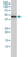
      Detection of human TARDBP by immunoblotting. Sample: Whole cell lysate from A431 cells. Primary antibody: Anti-TARDBP Mouse mAb (2E2-D3) (Cat. No. DR1075) (1-5 µg/ml). Detection: chemiluminescence.

      Detection of human TARDBP by staining paraffin sections. Sample: Formalin-fixed, paraffin-embedded human leiomyosarcoma tissue. Primary antibody: Anti-TARDBP Mouse mAb (2E2-D3) (Cat. No. DR1075) (5 µg/ml). Detection: colorimetric (DAB).

      Detection of human TARDBP by immunocytochemistry. Sample: HeLa cells fixed in paraformaldahyde. Primary antibody: Anti-TARDBP Mouse mAb (2E2-D3) (Cat. No. DR1075) (5 µg/ml). Detection: fluorescence.
      References
      ReferencesIlieva, E,V., et al. 2010. Free Radic. Biol. Med. 48, 1302.
      Wils H, et al. 2010. Proc. Natl. Acad. Sci. U S A 107 3858.
      Foulds, P,G., et al. 2009. Acta Neuropathol. 118 647.
      Product Information
      FormLiquid
      FormulationIn PBS, pH 7.7.
      Negative control293T cells
      Positive controlA431 cells, Human leiomyosarcoma tissue, HeLa cells
      PreservativeNone
      Quality LevelMQ100
      Applications
      Key Applications Immunoblotting (Western Blotting)
      Immunocytochemistry
      Paraffin Sections
      Application NotesImmunoblotting (1-5 µg/ml)
      Immunocytochemistry (10 µg/ml)
      Paraffin Sections (5 µg/ml)
      Application CommentsVariables associated with assay conditions will dictate the proper working dilution.
      Biological Information
      ImmunogenFull-length, recombinant human TARDBP (aa 1-261), expressed as a GST fusion protein.
      Clone2E2-D3
      HostMouse
      IsotypeIgG₁
      Species Reactivity
      • Human
      Antibody TypeMonoclonal Antibody
      Physicochemical Information
      Dimensions
      Materials Information
      Toxicological Information
      Safety Information according to GHS
      Safety Information
      Product Usage Statements
      Storage and Shipping Information
      Ship Code Blue Ice Only
      Toxicity Regulatory Review
      Storage -20°C
      Avoid freeze/thaw Avoid freeze/thaw
      Do not freeze Ok to freeze
      Special InstructionsFollowing initial thaw, aliquot and freeze (-20°C).
      Packaging Information
      Transport Information
      Supplemental Information
      Specifications
      Global Trade Item Number
      カタログ番号 GTIN
      DR1075-100UGCN 04055977226058

      Documentation

      Anti-TARDBP Mouse mAb (2E2-D3) (M)SDS

      タイトル

      英語版製品安全データシート((M)SDS) 

      参考資料

      参考資料の概要
      Ilieva, E,V., et al. 2010. Free Radic. Biol. Med. 48, 1302.
      Wils H, et al. 2010. Proc. Natl. Acad. Sci. U S A 107 3858.
      Foulds, P,G., et al. 2009. Acta Neuropathol. 118 647.
      データシート

      Note that this data sheet is not lot-specific and is representative of the current specifications for this product. Please consult the vial label and the certificate of analysis for information on specific lots. Also note that shipping conditions may differ from storage conditions.

      Revision04-November-2010 RFH
      SynonymsAnti-TAR DNA Binding Protein
      ApplicationImmunoblotting (1-5 µg/ml)
      Immunocytochemistry (10 µg/ml)
      Paraffin Sections (5 µg/ml)
      Application Data
      Detection of human TARDBP by immunoblotting. Sample: Whole cell lysate from A431 cells. Primary antibody: Anti-TARDBP Mouse mAb (2E2-D3) (Cat. No. DR1075) (1-5 µg/ml). Detection: chemiluminescence.

      Detection of human TARDBP by staining paraffin sections. Sample: Formalin-fixed, paraffin-embedded human leiomyosarcoma tissue. Primary antibody: Anti-TARDBP Mouse mAb (2E2-D3) (Cat. No. DR1075) (5 µg/ml). Detection: colorimetric (DAB).

      Detection of human TARDBP by immunocytochemistry. Sample: HeLa cells fixed in paraformaldahyde. Primary antibody: Anti-TARDBP Mouse mAb (2E2-D3) (Cat. No. DR1075) (5 µg/ml). Detection: fluorescence.
      DescriptionPurified mouse monoclonal antibody. Recognizes the ~45 kDa TARDBP protein.
      BackgroundHIV-1, the causative agent of acquired immunodeficiency syndrome (AIDS), contains an RNA genome that produces a chromosomally integrated DNA during the replicative cycle. Activation of HIV-1 gene expression by the transactivator Tat is dependent on an RNA regulatory element (TAR) located downstream of the transcription initiation site. TARDBP is a transcriptional repressor that binds to chromosomally integrated TAR DNA and represses HIV-1 transcription.
      HostMouse
      ImmunogenFull-length, recombinant human TARDBP (aa 1-261), expressed as a GST fusion protein.
      Clone2E2-D3
      IsotypeIgG₁
      Specieshuman
      Positive controlA431 cells, Human leiomyosarcoma tissue, HeLa cells
      Negative control293T cells
      FormLiquid
      FormulationIn PBS, pH 7.7.
      PreservativeNone
      CommentsVariables associated with assay conditions will dictate the proper working dilution.
      Storage Avoid freeze/thaw
      -20°C
      Do Not Freeze Ok to freeze
      Special InstructionsFollowing initial thaw, aliquot and freeze (-20°C).
      Toxicity Regulatory Review
      ReferencesIlieva, E,V., et al. 2010. Free Radic. Biol. Med. 48, 1302.
      Wils H, et al. 2010. Proc. Natl. Acad. Sci. U S A 107 3858.
      Foulds, P,G., et al. 2009. Acta Neuropathol. 118 647.