


AP1168 Sigma-AldrichAnti-TNFSF14 Mouse mAb (4E3)

This Anti-TNFSF14 Mouse mAb (4E3) is validated for use in ELISA, Immunoblotting, Paraffin Sections for the detection of TNFSF14.
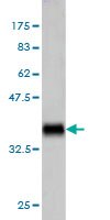
More>> This Anti-TNFSF14 Mouse mAb (4E3) is validated for use in ELISA, Immunoblotting, Paraffin Sections for the detection of TNFSF14. Less<<Anti-TNFSF14 Mouse mAb (4E3) MSDS (material safety data sheet) or SDS, CoA and CoQ, dossiers, brochures and other available documents.
Synonyms: Anti-Tumor Necrosis Factor Receptor Like 2, Anti-Tumor Necrosis Factor Superfamily Member 14
Recommended Products
Overview
| Replacement Information |
|---|
Key Spec Table
| Species Reactivity | Host | Antibody Type |
|---|---|---|
| H | M | Monoclonal Antibody |
Pricing & Availability
| Catalogue Number | Availability | Packaging | Qty/Pack | Price | Quantity | |
|---|---|---|---|---|---|---|
| AP1168-100UGCN |
|
100 μg |
|
— |


| References |
|---|
| Product Information | |
|---|---|
| Form | Liquid |
| Formulation | In PBS, pH 7.2. |
| Positive control | Human spleen tissue |
| Preservative | None |
| Quality Level | MQ100 |
| Physicochemical Information |
|---|
| Dimensions |
|---|
| Materials Information |
|---|
| Toxicological Information |
|---|
| Safety Information according to GHS |
|---|
| Safety Information |
|---|
| Product Usage Statements |
|---|
| Packaging Information |
|---|
| Transport Information |
|---|
| Supplemental Information |
|---|
| Specifications |
|---|
| Global Trade Item Number | |
|---|---|
| Catalogue Number | GTIN |
| AP1168-100UGCN | 04055977212693 |
Documentation
Anti-TNFSF14 Mouse mAb (4E3) SDS
| Title |
|---|