コンテンツへスキップ
Merck
ホーム3D細胞培養Millicell®ハンギングセルカルチャーインサートを用いたバリア形成および透過性アッセイ

Millicell®ハンギングセルカルチャーインサートを用いたバリア形成および透過性アッセイ

バリアアッセイの概要

培養細胞のバリアアッセイは、薬物送達、薬物吸収、創傷治癒、細菌/ウイルス感染、炎症などの研究においてバリア組織(腎臓、肺、腸など)をモデル化するために利用されています。上皮細胞を単一細胞懸濁液として播種し、融合して一つの組織になるまで成長させることで、これらのバリアがin vitro で形成されます。通常、この組織は一層の上皮細胞の厚さで、「上皮単層」と呼ばれます。標準的なバリアアッセイでは、細胞を多孔膜に播種し、経上皮電気抵抗(TEER)測定を介して非侵襲的に観察します。TEER測定は、評価や実験の前に上皮単層バリア形成を示すために、検証済みでラベリング不要、かつ迅速な測定技術です。バリア形成は、抵抗値の増加によって観察できます。上皮単層が密になると、より高い抵抗値でプラトーに達します。ほとんどのバリアアッセイは、単層上皮細胞の細胞バリアタイプです。肺から血液、腎臓でのろ過、または腸の吸収における単一細胞の厚さをモデル化し、上皮細胞間の細胞間隙空間を分析するために使用されます。 細胞バリアの完全性を確認した後、これらの組織はバリアの透過性、破壊、または強度を調べる実験に使用されます。

タイトジャンクションの”孔”は、上皮組織における細胞間隙輸送の生理的ゲートキーパーです。細胞間隙透過性アッセイは、特定の時間内に上皮細胞単層を通過する分子の量を測定することによって、単層バリア形成と細胞間隙タイトジャンクションの密度を評価します。このタイプのアッセイでは生きた細胞が必要で、通常は非破壊的アッセイが用いられます(例:ルシフェルイエロー細胞間隙透過性アッセイ)。免疫染色および組織免疫細胞化学(ICC)は、電子顕微鏡を使用しなくても良いようにするためによく用いられる方法です。これらの分析方法の多くは、染色試薬、薬物複合体、または移動したターゲットタンパク質の量として追跡し、バリアの通過を見るための方法です。代替分析方法としては、クリスタルバイオレットや蛍光抗体染色、ICCなどの染色法を使用して膜を可視化します。
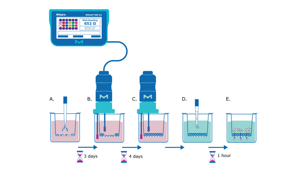
Millicell®ハンギングセルカルチャーインサートを使用したバリアアッセイの原理。細胞をインサートに播種し、3日後にMillicell 3.0 ERSを使用してTEER測定を行います。播種後 7 日で TEER 測定値が安定し、培地を交換して透過性アッセイの浅酌試薬を添加できるようになります。小分子を添加して単層の細胞とともに培養し、その通過を測定できます。

図1.Millicell®ハンギングセルカルチャーインサートを使用したバリアアッセイの原理。A) 0日目に、上皮細胞が単一細胞懸濁液をMillicell®ハンギングセルカルチャーインサートメンブレンに播種します。B) 数日後(MDCK細胞の場合は播種から約3日後)、細胞は密に成長し、TEER測定を開始するのに十分なコンフルエンスに達し、抵抗値測定に基づいて上皮細胞単層の形成を評価します。C) TEER測定は、細胞が一貫した抵抗値でプラトーに達するまで、単層バリア形成の状態を追跡するために、毎日または隔日で行います(MDCK細胞の場合は播種から約7日後)。D) この時点で、培地をバリアアッセイ液(Hank’s バランス塩溶液)に交換し、透過性アッセイ用の染色試薬/分子(例:ルシフェルイエロー染色試薬)をメンブレンの上部側に添加します。E) 小分子は、プロトコルで推奨する時間(例:1時間)、37°Cで上皮単層とともに培養し、その後、インサートの基底側からの液体を分析し、対象分子の通過量を測定します。 

ルシファーイエローアッセイを行うために必要なもの

  • 0.4または1 µmの孔サイズのMillicell®ハンギングセルカルチャーインサート(以下の「Millicell® ハンギングセルカルチャーインサートの選択」を参照)

Millicell®ハンギングセルカルチャーインサートの選択法

多孔膜は標準的なポリスチレン膜表面に比べて、2.5D細胞培養において大幅に改善された特性を有するため、バリアアッセイはハンギングインサートを用いて行います。インサートのメンブレンは、アッセイによってポアサイズが異なります。このことは異なったアッセイを行う際に重要となります。たとえば、遊走アッセイでは、実験に用いる細胞のタイプによって、最も広範なポアサイズから選択することになります。

TEER測定方法

Millicell®ERS 3.0 電圧抵抗計は、低電流と電圧を使用して、細胞培養における上皮単層のコンフルエンシーを非破壊的に測定します。TEER機器は、低AC電流を生成し、高いDC電流によって引き起こされるであろう電極金属の堆積や組織への悪影響を回避します。細胞単層のコンフルエンシーは、TEER測定機を使用して測定する組織抵抗値の増加またはプラトーに達したかによって判断します。

MDCK細胞培養液

24ウェル Millicell®ハンギングセルカルチャーインサートへの細胞播種

  1. MDCK細胞を60~80%コンフルエントになるまで培養します。
  2. 培養フラスコから培地を吸引します。
  3. 無菌PBSを室温で3分間加えます。
  4. 培養フラスコから液体を吸引します。
  5. トリプシンを培養フラスコに加え、37°C、5% CO2の細胞培養インキュベーターで培養します。
  6. 完全細胞培養培地を等量加えることでトリプシン反応を停止します。
  7. 液体懸濁液を集め、300 x gで3分間遠心分離します。
  8. 細胞を1 mLの完全な細胞培養培地に再懸濁します(上記のMDCK細胞培養培地のレシピを参照)。
  9. トリパンブルーヘモサイトメーターを使用して細胞をカウントします。90%のトリパンブルーと10%の細胞懸濁液を混合します(例:180 µLのトリパンブルーと20 µLの細胞懸濁液)。
  10. ヘモサイトメーターで生細胞をカウントし、ミリリットルあたりの細胞数を計算します。
  11. 24ウェルのメンブレンの基底側に900 µLの完全培地を加えます。
  12. 適切な体積に細胞懸濁液を再懸濁し、希望する細胞播種密度(例:1,250細胞/インサート、2,500細胞/インサート、5,000細胞/インサート)を含む200 µLの懸濁液を分注します。
  13. 37°C、5%二酸化炭素の細胞培養インキュベーターに静置し、隔日で培地を交換します。
    注記:培地をMillicell®ハンギングセルカルチャーインサートとレシーバープレートに加える前に、完全培地を予め温めておくことを推奨します。

細胞の抵抗値測定

  1. プレートが室温に達するまで放置します(15~30分)。
  2. プレートマップをロードし、Millicell® ERS 3.0デジタル電圧抵抗計でウェルをグループに割り当てます(ユーザーガイドのp. 11を参照)。
  3. 検証アダプターを使用してMillicell® ERS 3.0デジタル電圧抵抗計を検証します( ユーザーガイドのp. 22を参照)。
  4. MODEスイッチをOhmsに設定し、電源スイッチをオンにします。
  5. 電極を浸し、短い方の先端がMillicell®ハンギングセルカルチャーインサートの上部側に、長い方の先端が外側のウェルに入るようにします。短い方の先端はメンブレン上で成長する細胞に接触してはいけません。長い方の先端は外側のウェルの底に軽く触れるくらいに設定します。安定して再現性のある結果を得るために、電極がプレートおよびインサートの底に対して90°の角度で安定して保持されていることを確認してください。
    注記:測定間でのサンプルの持ち越し(キャリーオーバー)を防ぐために洗浄が必要な場合は、蒸留水ではなく細胞培養液を使用してください。
  6. 抵抗値を記録します。
  7. 空のウェル/細胞培養インサート(例:細胞と完全培地のないセルカルチャーインサート)に培養液または電解質溶液を加えることで、ブランク抵抗を測定します。
  8. ブランクインサート全体の抵抗値を測定し、この値を「ゼロ」または「バックグラウンド」抵抗レベルとして使用します。真の組織の抵抗値を得るためには、ブランクインサートの抵抗値を組織(細胞を培養しているセルカルチャーインサート)の抵抗値から引きます。
  9. 抵抗は組織の面積に反比例します。膜が大きいほど、抵抗値は低くなります。
    単位面積抵抗 (Ω × cm2) = 抵抗 (Ω) ×有効膜面積 (cm2)
    注記:抵抗はサンプルの温度、pH、および溶液中の電極の深さによって変動する可能性があります。

TEERアプリケーションノート、ガイド、その他情報

ルシファーイエローバリアアッセイサンプルプロトコル

手順

  1. メンブレン/ウェルの上部および基底側から培地を吸引します。
    TIP:吸引中にメンブレンに触れたり、突いたりしないように注意してください。
  2. メンブレンとウェルを予め温めた Hank’sバランス塩溶液(HBSS)で洗浄/すすぎます。
    TIP:予め温めたHBSSは細胞単層への影響が最も少ないですが、PBSに使用はより有害で、染色試薬の通過率が高くなります。
  3. 上部および基底側からHBSSを吸引します。
    TIP:吸引中にメンブレンに触れたり、突いたりしないように注意してください。
  4. メンブレンの基底側に900 µLのHBSSを分注します。
  5. メンブレンの上部側に200 µLのルシフェルイエロー試薬 (100 µg/mL)を分注します。
    TIP:ルシフェルイエロー試薬を誤って基底側に添加したり、こぼしたりしないように特に注意してください。このことによってアッセイ結果の精度が損なわれます。ピペッティング時にメンブレンを突かないでください。
  6. 37°C、5% CO2インキュベーターに1時間置きます。ポアサイズの小さいメンブレン(例: 0.4 µmのクリアMillicell®インサート(PCHT24H48)の場合は、インキュベーション時間を2~3時間に延長します。 
    TIP:最良の結果を得るために、プレートを水平に保ち、振動させないでください。
  7. この時間を利用して、サンプル値を特定するためのアッセイプレート用の標準を作成します。
        a.  100%標準 [Std(100)] を作成します:
             200 µLのルシフェルイエロー試薬(100 µg/mL)と900 µLのHBSSを混合します。
        b.  標準を1:1で希釈し、希釈系列を作ります。
             500 µLのStd(100)と500 µLのHBSSを混合するとStd(50)が得られます。
        c.  最も濃度の低い標準(100% HBSSを除く)は0.39%のルシフェルイエローを含むことになります(これにより9つの標準とHBSS
             の「ゼロ」の標準ができます)。
        d.  各標準の100 µLを蛍光分光プレートに追加します(3回繰り返し)。
  8. プレートをインキュベーターから取り出し、メンブレンの基底側から100 µLの液体を集め、蛍光分光法プレートに置きます。
    TIP:最良の結果を得るために、これを3回繰り返して、結果をテクニカルレプリケートとして平均化できるようにします。
  9. プレートリーダーで蛍光分光プレートを読み取り、485 nmの励起波長と535 nmの放出波長を使用して蛍光吸収を測定します。
  10. 生成された標準曲線を使用して、各ウェルのルシフェルイエロー濃度を計算します。これらの値は、各サンプルのパススルー率を決定するために使用できます。
    RFU = relative fluorescence units(相対蛍光単位)
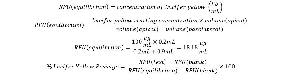
RFU平衡の公式
  1. 通常、ルシフェルイエロー透過性アッセイの成功基準は、5%未満のパススルーに設定されています。

バリアアッセイデータ例

Millicell®ハンギング細胞培養インサートを用いたMDCKバリア形成アッセイ

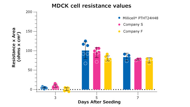
図3.すべてのメンブレンは、高密度(1×10⁸)0.4 µmポアサイズのPET 24ウェルメンブレンです。これらの結果は、Millicell®ハンギングセルカルチャーインサート(PTHT24H48)と2つのブランドのインサートを比較しています。MDCK細胞は、すべての3つのブランドで同じ播種密度で並行して播種しました。播種から7日後の3つのブランドのインサートは、互いに有意な差がなく、単層形成の類似した動態を示しています。
Millicell®ハンギングセルカルチャーインサートを用いたルシフェルイエロー透過性アッセイ

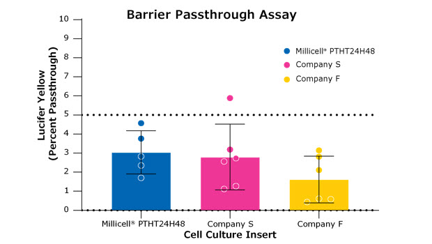
図4.ルシフェルイエロー透過性アッセイでは、細胞単層の形成と細胞間のタイトジャンクションを試験します。すべてのメンブレンは、高密度(1 x 10⁸孔)0.4 µmポアサイズのPETメンブレンで、24ウェルフォーマットです。これらの結果は、Millicell®ハンギングセルカルチャーインサート(PTHT24H48)と他の2つのブランドのインサートを比較しています。このアッセイは、図3のアッセイの7日目に実施しました。バリア形成は、メンブレンの上部側から基底側に染色試薬の5%未満の通過がある場合に成功と見なされます(通過は図1の最終パネルに示されています)。これらのインサート間に統計的な差はありませんでした。
Millicell® ERS-2 電圧抵抗計を使用したバリアの完全性TEER 測定

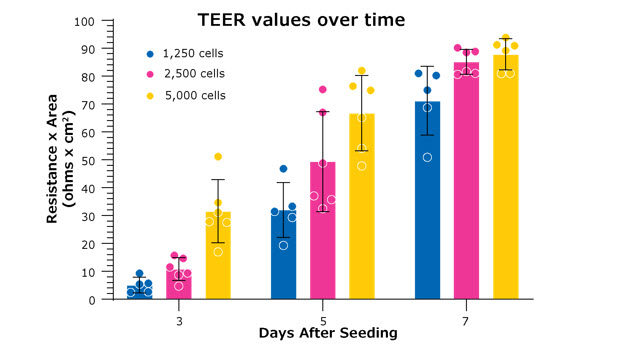
図5.TEER値は、Millicell®ハンギングセルカルチャーインサート上の細胞のコンフルエンスと相関する初期播種密度に依存します。より高い細胞播種密度では、細胞単層がより早く形成されます。実験、研究目的によっては、より高いまたは低い播種密度であることがより有益である場合があります。上記の例では、MDCK細胞は同じストックから播種され、指定された日に抵抗を測定しました。すべての測定は、Millicell® ERS 3.0デジタル電圧抵抗計とMillicell® ハンギングセルカルチャーインサート(PTHT24H48)を使用しました。 

バリアアッセイのヒントとコツ

  • 細胞タイプとアッセイに最適なメンブレンを選択してください(上記の表1 を参照)。
  • 細胞が十分な栄養を得て、単層/タイトジャンクション形成のための重要な細胞シグナルを維持するために、2日に1回培地を交換してください。
  • アッセイの前に細胞をインキュベーターの外に長時間放置しないでください。ルシフェルイエロー透過性アッセイの結果や他の類似のアッセイに悪影響を及ぼす可能性があります。
  • TEERプローブの電極が培地に完全に浸されていることを確認し、最も安定した測定値を得てください。
  • 一貫性のある比較可能な測定値を得るために、各ウェル/インサートでTEERプローブを同じ深さで保持してください。
  • 安定して再現性のある結果を得るために、電極がプレートインサートに対して90°の角度で配置されていることを確認してください。
  • 抵抗値は温度によって変動します。TEER測定値は、低温(低°C)で増加(高いオームΩ)します。測定値を測定する前に、サンプルが室温になるのを待ってください。
  • 抵抗値は培地のpHに影響されます。インキュベーターからすぐに引き出されたサンプルには、より高い二酸化炭素含量で、これは低い抵抗値に関連します。二酸化炭素の量は、室温に適応する際に大気中の二酸化炭素と平衡化します。
  • TEER電極は、細胞培養培地からのタンパク質の蓄積を避けるために、定期的にTergazymeなどの酵素洗剤で洗浄してください。この洗浄手順は、Millicell® ERS 3.0ユーザーガイドに記載されています。
  • Millicell® ERS 3.0のケーブルは、電波からの干渉に脆弱です。これらの電波は、携帯電話、ノートパソコン、コンピュータ、放射線機器(MRI、X線など)、さらにはスマートウォッチから発生します。実験エリアを、これらの干渉が最も少ない場所にすることを検討してください。
  • Millicell® ERS 3.0電圧抵抗計またはその電極プローブをUV光の下に置かないでください。電極センサーが損傷し、不正確で変動のある結果、または測定できない原因になります。
  • 測定間でのサンプルのキャリーオーバーを防ぐために洗浄が必要な場合は、蒸留水ではなく細胞培養液を使用してください。
  • 吸引中にメンブレンに触れたり、突いたりしないように注意してください。
  • ルシフェルイエロー透過性アッセイ中は、予め温めたHBSSが細胞単層への影響が最も少なくなります。PBSでは染色試薬の通過率が高くなります。
  • ルシフェルイエロー試薬を誤って基底側に添加したり、こぼしたりしないように特に注意してください。これによりアッセイ結果の精度が損なわれます。ピペッティング時にメンブレンを突かないでください。
  • ルシフェルイエロー試薬をメンブレンに加えたら、最良の結果を得るためにプレートを水平に保ち、振動させないでください。
  • ルシフェルイエロー透過性アッセイ終了時に、基底側の液体を混合した後に収集(上下にピペッティング)し、最良の結果を得るために通過結果の測定を3回繰り返してください。

関連製品

Loading

参考文献

1.
Sawamiphak S, Ritter M, Acker-Palmer A. 2010. Preparation of retinal explant cultures to study ex vivo tip endothelial cell responses. Nat Protoc. 5(10):1659-1665. https://doi.org/10.1038/nprot.2010.130
2.
Tamari T, Kawar-Jaraisy R, Doppelt O, Giladi B, Sabbah N, Zigdon-Giladi H. The Paracrine Role of Endothelial Cells in Bone Formation via CXCR4/SDF-1 Pathway. Cells. 9(6):1325. https://doi.org/10.3390/cells9061325
3.
Hamdollah Zadeh M, Glass CA, Magnussen A, Hancox JC, Bates DO. 2008. VEGF-Mediated Elevated Intracellular Calcium and Angiogenesis in Human Microvascular Endothelial CellsIn Vitroare Inhibited by Dominant Negative TRPC6. Microcirculation. 15(7):605-614. https://doi.org/10.1080/10739680802220323
4.
Hadjichristou C, Papachristou E, Bonovolias I, Bakopoulou A. 2020. Three-dimensional tissue engineering-based Dentin/Pulp tissue analogue as advanced biocompatibility evaluation tool of dental restorative materials. Dental Materials. 36(2):229-248. https://doi.org/10.1016/j.dental.2019.11.013
5.
DU H, SHI H, CHEN D, ZHOU Y, CHE G. 2015. Cross-talk between endothelial and tumor cells via basic fibroblast growth factor and vascular endothelial growth factor signaling promotes lung cancer growth and angiogenesis. 9(3):1089-1094. https://doi.org/10.3892/ol.2015.2881
6.
Del Tatto M, Ng T, Aliotta JM, Colvin GA, Dooner MS, Berz D, Dooner GJ, Papa EF, Hixson DC, Ramratnam B, et al. 2011. Marrow cell genetic phenotype change induced by human lung cancer cells. Experimental Hematology. 39(11):1072-1080. https://doi.org/10.1016/j.exphem.2011.08.008
7.
Hoeks C, Vanheusden M, Peeters LM, Stinissen P, Broux B, Hellings N. Treg-Resistant Cytotoxic CD4+ T Cells Dictate T Helper Cells in Their Vicinity: TH17 Skewing and Modulation of Proliferation. IJMS. 22(11):5660. https://doi.org/10.3390/ijms22115660
8.
Sygnecka K, Heider A, Scherf N, Alt R, Franke H, Heine C. 2015. Mesenchymal Stem Cells Support Neuronal Fiber Growth in an Organotypic Brain Slice Co-Culture Model. Stem Cells and Development. 24(7):824-835. https://doi.org/10.1089/scd.2014.0262
9.
Maeda T, Ibi M, Shimazu S, Akaike A. 1998. Co-culture With the Striatum Attenuates N-Methyl-D-aspartate Cytotoxicity in Dopaminergic Neurons of Rat Mesencephalic Slice Cultures. Japanese Journal of Pharmacology. 77(2):161-168. https://doi.org/10.1254/jjp.77.161
10.
Whitman MC, Bell JL, Nguyen EH, Engle EC. Ex Vivo Oculomotor Slice Culture from Embryonic GFP-Expressing Mice for Time-Lapse Imaging of Oculomotor Nerve Outgrowth. JoVE.(149): https://doi.org/10.3791/59911
11.
Robinson J, Turkington S, Abey S, Kenea N, Henderson W. 2019. Differential gene expression and gene-set enrichment analysis in Caco-2 monolayers during a 30-day timeline with Dexamethasone exposure. Tissue Barriers. 7(3):e1651597. https://doi.org/10.1080/21688370.2019.1651597
12.
Ricciardolo FL, Folkerts G, Folino A, Mognetti B. 2018. Bradykinin in asthma: Modulation of airway inflammation and remodelling. European Journal of Pharmacology. 827181-188. https://doi.org/10.1016/j.ejphar.2018.03.017
13.
Vis MAM, Ito K, Hofmann S. Impact of Culture Medium on Cellular Interactions in in vitro Co-culture Systems. Front. Bioeng. Biotechnol.. 8 https://doi.org/10.3389/fbioe.2020.00911
14.
Matsushita T, Kibayashi T, Katayama T, Yamashita Y, Suzuki S, Kawamata J, Honmou O, Minami M, Shimohama S. 2011. Mesenchymal stem cells transmigrate across brain microvascular endothelial cell monolayers through transiently formed inter-endothelial gaps. Neuroscience Letters. 502(1):41-45. https://doi.org/10.1016/j.neulet.2011.07.021
15.
Kidney JC, Proud D. 2000. Neutrophil Transmigration across Human Airway Epithelial Monolayers. Am J Respir Cell Mol Biol. 23(3):389-395. https://doi.org/10.1165/ajrcmb.23.3.4068
16.
Van Oostveldt K, Paape M, Burvenich C. 2002. Apoptosis of Bovine Neutrophils Following Diapedesis Through a Monolayer of Endothelial and Mammary Epithelial Cells. Journal of Dairy Science. 85(1):139-147. https://doi.org/10.3168/jds.s0022-0302(02)74062-9
17.
Heise R, Vetter-Kauczok CS, Skazik C, Czaja K, Marquardt Y, Lue H, Merk HF, Bernhagen J, Baron JM. 2012. Expression and Function of Macrophage Migration Inhibitory Factor in the Pathogenesis of UV-Induced Cutaneous Nonmelanoma Skin Cancer?. 88(5):1157-1164. https://doi.org/10.1111/j.1751-1097.2012.01108.x
18.
Yu J, Kim HM, Kim KP, Son Y, Kim M, Park K. 2019. Ceramide kinase regulates the migration of bone marrow-derived mesenchymal stem cells. Biochemical and Biophysical Research Communications. 508(2):361-367. https://doi.org/10.1016/j.bbrc.2018.11.154
19.
Wang X, Ma G, Zhang R, Liu L, Zhu J, Zhu H. 2019. Molecular characterization and biological functioning of interleukin-8 in Siberian sturgeon (Acipenser baeri). Fish & Shellfish Immunology. 9091-101. https://doi.org/10.1016/j.fsi.2019.04.010
20.
Trost B, Walker S, Wang Z, Thiruvahindrapuram B, MacDonald JR, Sung WW, Pereira SL, Whitney J, Chan AJ, Pellecchia G, et al. 2018. A Comprehensive Workflow for Read Depth-Based Identification of Copy-Number Variation from Whole-Genome Sequence Data. The American Journal of Human Genetics. 102(1):142-155. https://doi.org/10.1016/j.ajhg.2017.12.007
21.
Spary LK, Salimu J, Webber JP, Clayton A, Mason MD, Tabi Z. 2014. Tumor stroma-derived factors skew monocyte to dendritic cell differentiation toward a suppressive CD14+PD-L1+phenotype in prostate cancer. OncoImmunology. 3(9):e955331. https://doi.org/10.4161/21624011.2014.955331
22.
Welch HC, Condliffe AM, Milne LJ, Ferguson GJ, Hill K, Webb LM, Okkenhaug K, Coadwell WJ, Andrews SR, Thelen M, et al. 2005. P-Rex1 Regulates Neutrophil Function. Current Biology. 15(20):1867-1873. https://doi.org/10.1016/j.cub.2005.09.050
23.
Zhang Y, Wang B, Meng X, Sun G, Gao C. 2011. Influences of Acid-Treated Multiwalled Carbon Nanotubes on Fibroblasts: Proliferation, Adhesion, Migration, and Wound Healing. Ann Biomed Eng. 39(1):414-426. https://doi.org/10.1007/s10439-010-0151-y
24.
Sokolowski JD, Chabanon-Hicks CN, Han CZ, Heffron DS, Mandell JW. Fractalkine is a â??find-meâ? signal released by neurons undergoing ethanol-induced apoptosis. Front. Cell. Neurosci.. 8 https://doi.org/10.3389/fncel.2014.00360
25.
Hagan K, Varelas P, Zheng H. 2022. Endocannabinoid System of the Blood?Brain Barrier: Current Understandings and Therapeutic Potentials. Cannabis and Cannabinoid Research. 7(5):561-568. https://doi.org/10.1089/can.2021.0101
26.
Tucci M, Ciavarella S, Strippoli S, Brunetti O, Dammacco F, Silvestris F. 2011. Immature dendritic cells from patients with multiple myeloma are prone to osteoclast differentiation in vitro. Experimental Hematology. 39(7):773-783.e1. https://doi.org/10.1016/j.exphem.2011.04.006
27.
Fell LH, Seiler-Mußler S, Sellier AB, Rotter B, Winter P, Sester M, Fliser D, Heine GH, Zawada AM. 2016. Impact of individual intravenous iron preparations on the differentiation of monocytes towards macrophages and dendritic cells. Nephrol. Dial. Transplant.. 31(11):1835-1845. https://doi.org/10.1093/ndt/gfw045
28.
Su Q, Igyártó BZ. 2019. Keratinocytes Share Gene Expression Fingerprint with Epidermal Langerhans Cells via mRNA Transfer. Journal of Investigative Dermatology. 139(11):2313-2323.e8. https://doi.org/10.1016/j.jid.2019.05.006
29.
Hertz J, Qu B, Hu Y, Patel RD, Valenzuela DA, Goldberg JL. 2014. Survival and Integration of Developing and Progenitor-Derived Retinal Ganglion Cells following Transplantation. Cell Transplant. 23(7):855-872. https://doi.org/10.3727/096368913x667024
30.
Shetty AK, Turner DA. 1999. Neurite Outgrowth from Progeny of Epidermal Growth Factor–Responsive Hippocampal Stem Cells is Significantly Less Robust than From Fetal Hippocampal Cells Following Grafting Onto Organotypic Hippocampal Slice Cultures: Effect of Brain-Derived Neurotrophic Factor. Journal of Neurobiology. https://doi.org/10.1002/(sici)1097-4695(19990215)38:3<391::aid-neu8>3.0.co;2-4
31.
Ichikawa-Tomikawa N, Ogawa J, Douet V, Xu Z, Kamikubo Y, Sakurai T, Kohsaka S, Chiba H, Hattori N, Yamada Y, et al. 2012. Laminin ?1 is essential for mouse cerebellar development. Matrix Biology. 31(1):17-28. https://doi.org/10.1016/j.matbio.2011.09.002
32.
Namsolleck P, Boato F, Schwengel K, Paulis L, Matho KS, Geurts N, Thöne-Reineke C, Lucht K, Seidel K, Hallberg A, et al. 2013. AT2-receptor stimulation enhances axonal plasticity after spinal cord injury by upregulating BDNF expression. Neurobiology of Disease. 51177-191. https://doi.org/10.1016/j.nbd.2012.11.008
33.
Lee D, Cho K. 2005. The effects of epidermal keratinocytes and dermal fibroblasts on the formation of cutaneous basement membrane in three-dimensional culture systems. Arch Dermatol Res. 296(7):296-302. https://doi.org/10.1007/s00403-004-0529-5
34.
Díaz-Coránguez M, Segovia J, López-Ornelas A, Puerta-Guardo H, Ludert J, Chávez B, Meraz-Cruz N, González-Mariscal L. Transmigration of Neural Stem Cells across the Blood Brain Barrier Induced by Glioma Cells. PLoS ONE. 8(4):e60655. https://doi.org/10.1371/journal.pone.0060655
35.
Huang S, Liu F, Niu Q, Li Y, Liu C, Zhang L, Ni D, Pu X. GLIPR-2 Overexpression in HK-2 Cells Promotes Cell EMT and Migration through ERK1/2 Activation. PLoS ONE. 8(3):e58574. https://doi.org/10.1371/journal.pone.0058574
36.
den Broeder A, Wanten G, Oyen W, Naber T, van Riel P, Barrera P. 2003. Neutrophil Migration and Production of Reactive Oxygen Species During Treatment with a Fully Human Anti-Tumor Necrosis Factor-Alpha Monoclonal Antibody in Patients with Rheumatoid Arthritis.  The Journal of Rheumatology. 30(2):232-237.
37.
Nishihara H, Soldati S, Mossu A, Rosito M, Rudolph H, Muller WA, Latorre D, Sallusto F, Sospedra M, Martin R, et al. 2020. Human CD4+ T cell subsets differ in their abilities to cross endothelial and epithelial brain barriers in vitro. Fluids Barriers CNS. 17(1): https://doi.org/10.1186/s12987-019-0165-2
38.
Zhang R, Wu Y, Zhao M, Liu C, Zhou L, Shen S, Liao S, Yang K, Li Q, Wan H. 2009. Role of HIF-1? in the regulation ACE and ACE2 expression in hypoxic human pulmonary artery smooth muscle cells. American Journal of Physiology-Lung Cellular and Molecular Physiology. 297(4):L631-L640. https://doi.org/10.1152/ajplung.90415.2008
39.
Ning Y, Sun Q, Dong Y, Xu W, Zhang W, Huang H, Li Q. 2011. Slit2-N inhibits PDGF-induced migration in rat airway smooth muscle cells: WASP and Arp2/3 involved. Toxicology. 283(1):32-40. https://doi.org/10.1016/j.tox.2011.01.026
40.
Wang Y, Wang HJ, Liao Y, Tsai P, Chen K, Cheng H, Lin R, Juo SH. 2012. MicroRNA-195 regulates vascular smooth muscle cell phenotype and prevents neointimal formation. 95(4):517-526. https://doi.org/10.1093/cvr/cvs223
41.
Zhang W, Tang B, Huang Q, Hua Z. 2013. Galangin inhibits tumor growth and metastasis of B16F10 melanoma. J. Cell. Biochem.. 114(1):152-161. https://doi.org/10.1002/jcb.24312
42.
Schittek B, Psenner K, Sauer B, Meier F, Iftner T, Garbe C. 2007. The increased expression of Y box-binding protein 1 in melanoma stimulates proliferation and tumor invasion, antagonizes apoptosis and enhances chemoresistance. Int. J. Cancer. 120(10):2110-2118. https://doi.org/10.1002/ijc.22512
43.
Meier F, Busch S, Lasithiotakis K, Kulms D, Garbe C, Maczey E, Herlyn M, Schittek B. 2007. Combined targeting of MAPK and AKT signalling pathways is a promising strategy for melanoma treatment. Br J Dermatol. 156(6):1204-1213. https://doi.org/10.1111/j.1365-2133.2007.07821.x
44.
Wang Y, Chang S, Wang C, Huang H, Tzeng S. 2020. The selective lipoprotein-associated phospholipase A2 inhibitor darapladib triggers irreversible actions on glioma cell apoptosis and mitochondrial dysfunction. Toxicology and Applied Pharmacology. 402115133. https://doi.org/10.1016/j.taap.2020.115133
45.
Bajetto A, Barbieri F, Dorcaratto A, Barbero S, Daga A, Porcile C, Ravetti JL, Zona G, Spaziante R, Corte G, et al. 2006. Expression of CXC chemokine receptors 1?5 and their ligands in human glioma tissues: Role of CXCR4 and SDF1 in glioma cell proliferation and migration. Neurochemistry International. 49(5):423-432. https://doi.org/10.1016/j.neuint.2006.03.003
46.
Luo L, Zhang L, Duan C, Wang B, He N, Abulimiti P, Lin Y. 2017. The inhibition role of miR-22 in hepatocellular carcinoma cell migration and invasion via targeting CD147. Cancer Cell Int. 17(1): https://doi.org/10.1186/s12935-016-0380-8
47.
Li J, Zhang S, Chen J, Du T, Wang Y, Wang Z. 2009. Modeled microgravity causes changes in the cytoskeleton and focal adhesions, and decreases in migration in malignant human MCF-7 cells. Protoplasma. 238(1-4):23-33. https://doi.org/10.1007/s00709-009-0068-1
48.
Baumann L, Prokoph S, Gabriel C, Freudenberg U, Werner C, Beck-Sickinger AG. 2012. A novel, biased-like SDF-1 derivative acts synergistically with starPEG-based heparin hydrogels and improves eEPC migration in vitro. Journal of Controlled Release. 162(1):68-75. https://doi.org/10.1016/j.jconrel.2012.04.049
49.
CADET ER, GAFNI RI, MCCARTHY EF, MCCRAY DR, BACHER JD, BARNES KM, BARON J. 2003. MECHANISMS RESPONSIBLE FOR LONGITUDINAL GROWTH OF THE CORTEX. The Journal of Bone and Joint Surgery-American Volume. 85(9):1739-1748. https://doi.org/10.2106/00004623-200309000-00013
50.
Zhao F, Li L, Guan L, Yang H, Wu C, Liu Y. 2014. Roles for GP IIb/IIIa and ?v?3 integrins in MDA-MB-231 cell invasion and shear flow-induced cancer cell mechanotransduction. Cancer Letters. 344(1):62-73. https://doi.org/10.1016/j.canlet.2013.10.019
51.
Jiang R, Zhang C, Liu G, Gu R, Wu H. 2017. Inhibits Proliferation, Migration and Invasion in Osteosarcoma Cells by Targeting ROCK1. American Journal of Cancer Research. 7(1):88.
52.
Jia D, Niu Y, Li D, Liu Z. 2018. lncRNA C2dat1 Promotes Cell Proliferation, Migration, and Invasion by Targeting miR-34a-5p in Osteosarcoma Cells. oncol res. 26(5):753-764. https://doi.org/10.3727/096504017x15024946480113
53.
Zhang J, Yang W, Zhou Y, Xiang Y, Wang L, Hu W, Wang W. Baicalein inhibits osteosarcoma cell proliferation and invasion through the miR?183/Ezrin pathway. Mol Med Report. https://doi.org/10.3892/mmr.2018.9036
54.
Liu Y, Zhao F, Gu W, Yang H, Meng Q, Zhang Y, Yang H, Duan Q. 2009. The Roles of Platelet GPIIb/IIIa and ?v?3 Integrins during HeLa Cells Adhesion, Migration, and Invasion to Monolayer Endothelium under Static and Dynamic Shear Flow. Journal of Biomedicine and Biotechnology. 20091-9. https://doi.org/10.1155/2009/829243
55.
Toyoda K, Tanaka K, Nakagawa S, Thuy DHD, Ujifuku K, Kamada K, Hayashi K, Matsuo T, Nagata I, Niwa M. 2013. Initial Contact of Glioblastoma Cells with Existing Normal Brain Endothelial Cells Strengthen the Barrier Function via Fibroblast Growth Factor 2 Secretion: A New In Vitro Blood?Brain Barrier Model. Cell Mol Neurobiol. 33(4):489-501. https://doi.org/10.1007/s10571-013-9913-z
56.
Hu M, Wang C, Li W, Lu W, Bai Z, Qin D, Yan Q, Zhu J, Krueger BJ, Renne R, et al. A KSHV microRNA Directly Targets G Protein-Coupled Receptor Kinase 2 to Promote the Migration and Invasion of Endothelial Cells by Inducing CXCR2 and Activating AKT Signaling. PLoS Pathog. 11(9):e1005171. https://doi.org/10.1371/journal.ppat.1005171
57.
Coureuil M, Lécuyer H, Scott MG, Boularan C, Enslen H, Soyer M, Mikaty G, Bourdoulous S, Nassif X, Marullo S. 2010. Meningococcus Hijacks a ?2-Adrenoceptor/?-Arrestin Pathway to Cross Brain Microvasculature Endothelium. Cell. 143(7):1149-1160. https://doi.org/10.1016/j.cell.2010.11.035
58.
Almajed FS, Forsythe SJ. 2016. Cronobacter sakazakii clinical isolates overcome host barriers and evade the immune response. Microbial Pathogenesis. 9055-63. https://doi.org/10.1016/j.micpath.2015.11.014
59.
Tian X, Hu T, Zhang H, He L, Huang X, Liu Q, Yu W, He L, Yang Z, Zhang Z, et al. 2013. Subepicardial endothelial cells invade the embryonic ventricle wall to form coronary arteries. Cell Res. 23(9):1075-1090. https://doi.org/10.1038/cr.2013.83
60.
Monteiro IP, Shukla A, Marques AP, Reis RL, Hammond PT. 2015. Spray-assisted layer-by-layer assembly on hyaluronic acid scaffolds for skin tissue engineering. J. Biomed. Mater. Res.. 103(1):330-340. https://doi.org/10.1002/jbm.a.35178
61.
Vahav I, Broek LJ, Thon M, Monsuur HN, Spiekstra SW, Atac B, Scheper RJ, Lauster R, Lindner G, Marx U, et al. 2020. Reconstructed human skin shows epidermal invagination towards integrated neopapillae indicating early hair follicle formation in vitro. J Tissue Eng Regen Med. 14(6):761-773. https://doi.org/10.1002/term.3039
62.
Gottipati A, Elder SH. 2016. Composite Chitosan-Calcium Phosphate Scaffolds for Cartilage Tissue Engineering.83-97. https://doi.org/10.1007/978-81-322-2511-9_4
63.
Horváth L, Umehara Y, Jud C, Blank F, Petri-Fink A, Rothen-Rutishauser B. Engineering an in vitro air-blood barrier by 3D bioprinting. Sci Rep. 5(1): https://doi.org/10.1038/srep07974
64.
Karp PH, Moninger TO, Weber SP, Nesselhauf TS, Launspach JL, Zabner J, Welsh MJ. An In Vitro Model of Differentiated Human Airway Epithelia: Methods for Establishing Primary Cultures.115-137. https://doi.org/10.1385/1-59259-185-x:115
65.
Mohamed Ameer J, Venkatesan RB, Damodaran V, K.V. N, Arumugam S, Pallickaveedu Rajan Asari AK, Kasoju N. 2020. Fabrication of co-cultured tissue constructs using a dual cell seeding compatible cell culture insert with a clip-on scaffold for potential regenerative medicine and toxicological screening applications. Journal of Science: Advanced Materials and Devices. 5(2):207-217. https://doi.org/10.1016/j.jsamd.2020.04.004
66.
Velazquez OC, Snyder R, Liu Z, Fairman RM, Herlyn M. 2002. Fibroblast?dependent differentiation of human microvascular endothelial cells into capillary?like, three?dimensional networks. FASEB j.. 16(10):1316-1318. https://doi.org/10.1096/fj.01-1011fje
67.
Schacht K, Jüngst T, Schweinlin M, Ewald A, Groll J, Scheibel T. 2015. Biofabrication of Cell-Loaded 3D Spider Silk Constructs. Angew. Chem. Int. Ed.. 54(9):2816-2820. https://doi.org/10.1002/anie.201409846
68.
Tan H, Trier S, Rahbek UL, Dufva M, Kutter JP, Andresen TL. A multi-chamber microfluidic intestinal barrier model using Caco-2 cells for drug transport studies. PLoS ONE. 13(5):e0197101. https://doi.org/10.1371/journal.pone.0197101
69.
Artursson P. 1990. Epithelial Transport Of Drugs In Cell Culture. I: A Model For Studying The Passive Diffusion Of Drugs Over Intestinal Absorbtive (Caco-2) Cells. Journal of Pharmaceutical Sciences. 79(6):476-482. https://doi.org/10.1002/jps.2600790604
70.
Artursson P, Palm K, Luthman K. 2001. Caco-2 monolayers in experimental and theoretical predictions of drug transport1PII of original article: S0169-409X(96)00415-2. The article was originally published in Advanced Drug Delivery Reviews 22 (1996) 67?84.1. Advanced Drug Delivery Reviews. 46(1-3):27-43. https://doi.org/10.1016/s0169-409x(00)00128-9
71.
Bailey CA, Bryla P, Malick A. 1996. The use of the intestinal epithelial cell culture model, Caco-2, in pharmaceutical development. Advanced Drug Delivery Reviews. 22(1-2):85-103. https://doi.org/10.1016/s0169-409x(96)00416-4
72.
Kuhfeld MT, Stratford RE. 1996. In vitro measurement of drug transport using a new diffusion chamber compatible with Millicell® culture supports: Performance with caco-2 monolayers. International Journal of Pharmaceutics. 133(1-2):47-58. https://doi.org/10.1016/0378-5173(95)04415-9
73.
van Zeijl MJ, Matlin KS. 1990. Microtubule perturbation inhibits intracellular transport of an apical membrane glycoprotein in a substrate-dependent manner in polarized Madin-Darby canine kidney epithelial cells.. Cell Regul. 1(12):921-936. https://doi.org/10.1091/mbc.1.12.921
74.
HIROTANI Y, IKEDA K, KATO R, MYOTOKU M, UMEDA T, IJIRI Y, TANAKA K. 2008. Protective Effects of Lactoferrin against Intestinal Mucosal Damage Induced by Lipopolysaccharide in Human Intestinal Caco-2 Cells. YAKUGAKU ZASSHI. 128(9):1363-1368. https://doi.org/10.1248/yakushi.128.1363
75.
Trotta V, Pavan B, Ferraro L, Beggiato S, Traini D, Des Reis LG, Scalia S, Dalpiaz A. 2018. Brain targeting of resveratrol by nasal administration of chitosan-coated lipid microparticles. European Journal of Pharmaceutics and Biopharmaceutics. 127250-259. https://doi.org/10.1016/j.ejpb.2018.02.010
76.
Baghirov H, Karaman D, Viitala T, Duchanoy A, Lou Y, Mamaeva V, Pryazhnikov E, Khiroug L, de Lange Davies C, Sahlgren C, et al. Feasibility Study of the Permeability and Uptake of Mesoporous Silica Nanoparticles across the Blood-Brain Barrier. PLoS ONE. 11(8):e0160705. https://doi.org/10.1371/journal.pone.0160705
77.
Milatz S, Piontek J, Hempel C, Meoli L, Grohe C, Fromm A, Lee IM, El-Athman R, Günzel D. 2017. Tight junction strand formation by claudin-10 isoforms and claudin-10a/-10b chimeras. Ann. N.Y. Acad. Sci.. 1405(1):102-115. https://doi.org/10.1111/nyas.13393
78.
Hoy B, Löwer M, Weydig C, Carra G, Tegtmeyer N, Geppert T, Schröder P, Sewald N, Backert S, Schneider G, et al. 2010. Helicobacter pyloriHtrA is a new secreted virulence factor that cleaves E?cadherin to disrupt intercellular adhesion. EMBO Rep. 11(10):798-804. https://doi.org/10.1038/embor.2010.114
79.
Chen Z, Ma T, Huang C, Zhang L, Zhong J, Han J, Hu T, Li J. 2014. Efficiency of transcellular transport and efflux of flavonoids with different glycosidic units from flavonoids of Litsea coreana L. in a MDCK epithelial cell monolayer model. European Journal of Pharmaceutical Sciences. 5369-76. https://doi.org/10.1016/j.ejps.2013.12.010
80.
Volpe DA. 2008. Variability in Caco-2 and MDCK Cell-Based Intestinal Permeability Assays. Journal of Pharmaceutical Sciences. 97(2):712-725. https://doi.org/10.1002/jps.21010
81.
Chen R, Shen C, Xu Q, Liu Y, Li B, Huang C, Ma T, Meng X, Wu M, Li J. 2021. The permeability characteristics and interaction of main components from Si-Ni-San in a MDCK epithelial cell monolayer model. Xenobiotica. 51(2):239-248. https://doi.org/10.1080/00498254.2017.1359433
82.
Wang X, Valenzano MC, Mercado JM, Zurbach EP, Mullin JM. 2013. Zinc Supplementation Modifies Tight Junctions and Alters Barrier Function of CACO-2 Human Intestinal Epithelial Layers. Dig Dis Sci. 58(1):77-87. https://doi.org/10.1007/s10620-012-2328-8