
17-441
Rac1/Cdc42 Activation Assay Kit
The Rac1/Cdc42 Activation Assay provides an effective method for detecting Rac & Cdc42 activity in cell lysates.
동의어(들):
Rac1 activation assay
크기 선택
제품정보 (DICE 배송 시 비용 별도)
species reactivity
human, mouse, rat
manufacturer/tradename
Upstate®
technique(s)
activity assay: suitable, affinity binding assay: suitable (G-protein)
NCBI accession no.
UniProt accession no.
shipped in
wet ice
Quality Level
Gene Information
human ... RAC1(5879)
General description
Biochem/physiol Actions
Anti-Rac1, clone 23A8: Species Cross-reactivity: Human, mouse and rat. Other species cross-reactivity unknown.
Anti-cdc42 Species Cross-reactivity: Human, mouse, rat and dog. Other species cross-reactivity unknown.
Preparation Note
Analysis Note
Other Notes
Anti-Rac1, clone 23A8, Catalog #05-389: One vial containing 250 μg of protein G purified mouse IgG2b in 250 μL of storage buffer (0.1 M Tris-glycine, pH 7.4, 0.15 M NaCl, containing 0.05% sodium azide).
Anti-cdc42, (mouse monoclonal IgG1) , Catalog # 05-542: One vial containing 50 μg of purified mouse IgG1 in 200 μL of 50% storage buffer (20 mM sodium phosphate, pH 7.5, 150 mM NaCl, 1.5 mM sodium azide containing, 1 mg/mL BSA) and 50% glycerol.
Mg2+ Lysis/Wash Buffer, 5X, Catalog # 20-168 : Two vials, each vial containing 18 mL of 5X MLB: 125 mM HEPES, pH 7.5, 750 mM NaCl, 5% Igepal CA-630, 50 mM MgCl2, 5 mM EDTA and 10% glycerol.
100X GTPγS, 10mM, Catalog # 20-176: One vial containing 50 μL of 10 mM GTPγS, 100X stock, in 50 mM Tris-HCl, pH 7.8, non-hydrolyzable analog of GTP. Sufficient to label 5 mL of cell lysates.
100X GDP, 100mM, Catalog # 20-177: One vial containing 50 μL of 100 mM GDP, 100X stock, in 50 mM Tris-HCl, pH 7.8. GDP (Guanosine 5′-Diphosphate) for in vitro labeling of G-proteins in the inactive form. Sufficient to label 5ml of cell lysates.
Legal Information
Disclaimer

signalword
Danger
저장 등급
10 - Combustible liquids
hcodes
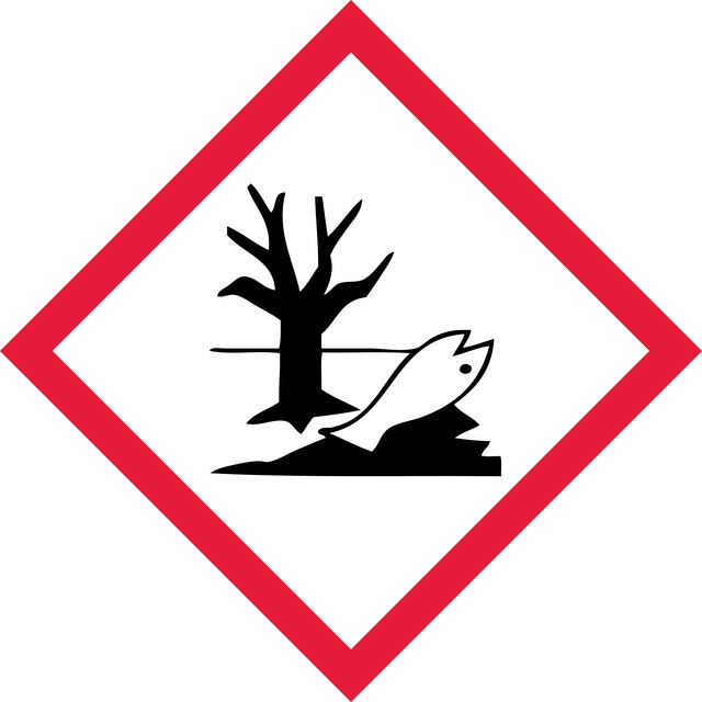
Hazard Classifications
Aquatic Acute 1 - Aquatic Chronic 2 - ED ENV 1 - Eye Dam. 1
wgk
WGK 3
시험 성적서(COA)
제품의 로트/배치 번호를 입력하여 시험 성적서(COA)을 검색하십시오. 로트 및 배치 번호는 제품 라벨에 있는 ‘로트’ 또는 ‘배치’라는 용어 뒤에서 찾을 수 있습니다.
관련 콘텐츠
"Aging: getting older, exhibiting the signs of age, the decline in the physical (and mental) well-being over time, leading to death. Since the beginning of time, man has been obsessed with trying to slow down, stop, or even reverse the signs of aging. Many have gone as far as experimenting with nutritional regimens, eccentric exercises, fantastic rituals, and naturally occurring or synthetic wonder-elements to evade the signs of normal aging. Biologically speaking, what is aging? And what does the latest research tell us about the possibility of discovering the elusive “fountain of youth”? Many advances in our understanding of aging have come from systematic scientific research, and perhaps it holds the key to immortality. Scientifically, aging can be defined as a systems-wide decline in organismal function that occurs over time. This decline occurs as a result of numerous events in the organism, and these events can be classified into nine “hallmarks” of aging, as proposed by López-Otin et al. (2013). Several of the pathologies associated with aging are a direct result of these events going to extremes and may also involve aberrant activation of proliferation signals or hyperactivity. The hallmarks of aging have been defined based on their fulfillment of specific aging related criteria, such as manifestation during normal aging, acceleration of aging if experimentally induced or aggravated, and retardation of aging if prevented or blocked, resulting in increased lifespan. The nine hallmarks of aging are genomic instability, telomere attrition, epigenetic alterations, loss of proteostasis, deregulated nutrient sensing, mitochondrial dysfunction, cellular senescence, stem cell exhaustion, and altered intercellular communication. The biological processes underlying aging are complex. By understanding the hallmarks in greater detail, we can get closer to developing intervention strategies that can make the aging process less of a decline, and more of a recline."
"Epigenetics describes heritable changes in gene expression caused by non-genetic mechanisms instead of by alterations in DNA sequence. These changes can be cell- or tissue-specific, and can be passed on to multiple generations. Epigenetic regulation enriches DNAbased information, allowing a cell to vary its response across diverse biological and environmental contexts. Although epigenetic mechanisms are primarily centered in the nucleus, these mechanisms can be induced by environmental signals such as hormones, nutrients, stress, and cellular damage, pointing to the involvement of cytoplasmic and extracellular factors in epigenetic regulation."
Signaling Product Guide: Antibodies, small molecule inhibitors, kits, assays and proteins for signaling research.
국제 무역 품목 번호
| SKU | GTIN |
|---|---|
| 17-441 | 04053252338700 |
자사의 과학자팀은 생명 과학, 재료 과학, 화학 합성, 크로마토그래피, 분석 및 기타 많은 영역을 포함한 모든 과학 분야에 경험이 있습니다..
고객지원팀으로 연락바랍니다.