


DR1075 Sigma-AldrichAnti-TARDBP Mouse mAb (2E2-D3)
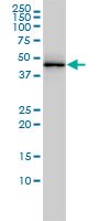

This Anti-TARDBP Mouse mAb (2E2-D3) is validated for use in Immunoblotting, Immunocytochemistry, Paraffin Sections for the detection of TARDBP.
More>> This Anti-TARDBP Mouse mAb (2E2-D3) is validated for use in Immunoblotting, Immunocytochemistry, Paraffin Sections for the detection of TARDBP. Less<<Anti-TARDBP Mouse mAb (2E2-D3) MSDS (material safety data sheet) or SDS, CoA and CoQ, dossiers, brochures and other available documents.
Synonyms: Anti-TAR DNA Binding Protein
Recommended Products
Overview
| Replacement Information |
|---|
Key Spec Table
| Species Reactivity | Host | Antibody Type |
|---|---|---|
| H | M | Monoclonal Antibody |
Products
| Catalogue Number | Packaging | Qty/Pack | |
|---|---|---|---|
| DR1075-100UGCN | 100 μg |


| References | |
|---|---|
| References | Ilieva, E,V., et al. 2010. Free Radic. Biol. Med. 48, 1302. Wils H, et al. 2010. Proc. Natl. Acad. Sci. U S A 107 3858. Foulds, P,G., et al. 2009. Acta Neuropathol. 118 647. |
| Product Information | |
|---|---|
| Form | Liquid |
| Formulation | In PBS, pH 7.7. |
| Negative control | 293T cells |
| Positive control | A431 cells, Human leiomyosarcoma tissue, HeLa cells |
| Preservative | None |
| Quality Level | MQ100 |
| Physicochemical Information |
|---|
| Dimensions |
|---|
| Materials Information |
|---|
| Toxicological Information |
|---|
| Safety Information according to GHS |
|---|
| Safety Information |
|---|
| Product Usage Statements |
|---|
| Packaging Information |
|---|
| Transport Information |
|---|
| Supplemental Information |
|---|
| Specifications |
|---|
| Global Trade Item Number | |
|---|---|
| Catalogue Number | GTIN |
| DR1075-100UGCN | 04055977226058 |
Documentation
Anti-TARDBP Mouse mAb (2E2-D3) MSDS
| Title |
|---|
References
| Reference overview |
|---|
| Ilieva, E,V., et al. 2010. Free Radic. Biol. Med. 48, 1302. Wils H, et al. 2010. Proc. Natl. Acad. Sci. U S A 107 3858. Foulds, P,G., et al. 2009. Acta Neuropathol. 118 647. |