
S7100
Kit de detección de la apoptosis in situ con peroxidasa ApopTag
The ApopTag Peroxidase In Situ Apoptosis Detection Kit detects apoptotic cells in situ by labeling & detecting DNA strand breaks by the TUNEL method.
Sinónimos:
Kit de ApopTag, Kit de detección de apoptosis in situ
Seleccione un Tamaño
Acerca de este artículo
Quality Level
manufacturer/tradename
ApopTag, Chemicon®
detection method
colorimetric
shipped in
dry ice
General description
Of all the aspects of apoptosis, the defining characteristic is a complete change in cellular morphology. As observed by electron microscopy, the cell undergoes shrinkage, chromatin margination, membrane blebbing, nuclear condensation and then segmentation, and division into apoptotic bodies which may be phagocytosed (11, 19, 24). The characteristic apoptotic bodies are short-lived and minute, and can resemble other cellular constituents when viewed by brightfield microscopy. DNA fragmentation in apoptotic cells is followed by cell death and removal from the tissue, usually within several hours (7). A rate of tissue regression as rapid as 25% per day can result from apparent apoptosis in only 2-3% of the cells at any one time (6). Thus, the quantitative measurement of an apoptotic index by morphology alone can be difficult.
DNA fragmentation is usually associated with ultrastructural changes in cellular morphology in apoptosis (26, 38). In a number of well-researched model systems, large fragments of 300 kb and 50 kb are first produced by endonucleolytic degradation of higher-order chromatin structural organization. These large DNA fragments are visible on pulsed-field electrophoresis gels (5, 43, 44). In most models, the activation of Ca2+- and Mg2+-dependent endonuclease activity further shortens the fragments by cleaving the DNA at linker sites between nucleosomes (3). The ultimate DNA fragments are multimers of about 180 bp nucleosomal units. These multimers appear as the familiar "DNA ladder" seen on standard agarose electrophoresis gels of DNA extracted from many kinds of apoptotic cells (e.g. 3, 7,13, 35, 44).
Another method for examining apoptosis via DNA fragmentation is by the TUNEL assay, (13) which is the basis of ApopTag technology. The DNA strand breaks are detected by enzymatically labeling the free 3′-OH termini with modified nucleotides. These new DNA ends that are generated upon DNA fragmentation are typically localized in morphologically identifiable nuclei and apoptotic bodies. In contrast, normal or proliferative nuclei, which have relatively insignificant numbers of DNA 3′-OH ends, usually do not stain with the kit. ApopTag Kits detect single-stranded (25) and double-stranded breaks associated with apoptosis. Drug-induced DNA damage is not identified by the TUNEL assay unless it is coupled to the apoptotic response (8). In addition, this technique can detect early-stage apoptosis in systems where chromatin condensation has begun and strand breaks are fewer, even before the nucleus undergoes major morphological changes (4, 8).
Apoptosis is distinct from accidental cell death (necrosis). Numerous morphological and biochemical differences that distinguish apoptotic from necrotic cell death are summarized in the following table (adapted with permission from reference 39).
Preparation Note
Precauciones
1.Los siguientes componentes del kit contienen cacodilato (ácido dimetilarsínico) de potasio como tampón: Tampón equilibrador (90416), tampón de reacción (90417) y enzima TdT (90418). Estos componentes son nocivos en caso de ingestión; evítese el contacto con los ojos y la piel (utilización de guantes y gafas) y lávense de inmediato las zonas de contacto.
2. Los conjugados de anticuerpos (90420) y las disoluciones de bloqueo (nº 10 y nº 13) contienen acida sódica al 0,08 % como conservante.
3. La enzima TdT (90418) contiene glicerol y no se congelará a -20°C. Para una máxima vida útil, no caliente este reactivo a temperatura ambiente antes de su dispensación.
Other Notes
Tampón de reacción 2,0 ml de -15°C a -25°C
Enzima TdT 0,64 ml de -15°C a -25°C
Tampón de parada/lavado 20 ml de -15°C a -25°C
Anti-digoxigenina-peroxidasa* 3,0 ml de 2°C a 8°C
Cubreobjetos de plástico 100 cada uno. Temperatura ambiente
Nota: Es necesaria la adquisición por separado de DAB (sustrato de la peroxidasa). No se suministra con este kit.
Número de muestras por kit: Se suministra material suficiente para teñir 40 muestras de tejido de aproximadamente 50 mm2 cada una cuando se utiliza según las instrucciones. El tampón de reacción se consumirá completamente antes que los demás reactivos cuando los kit se utilizan para muestras montadas en portaobjetos.
Legal Information


signalword
Danger
hcodes
Hazard Classifications
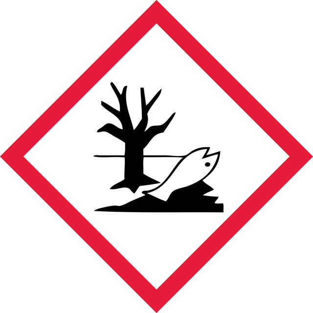
Aquatic Chronic 2 - Carc. 1B - Skin Sens. 1 - STOT RE 2 Inhalation
target_organs
Respiratory Tract
Clase de almacenamiento
6.1C - Combustible acute toxic Cat.3 / toxic compounds or compounds which causing chronic effects
wgk
WGK 3
Certificados de análisis (COA)
Busque Certificados de análisis (COA) introduciendo el número de lote del producto. Los números de lote se encuentran en la etiqueta del producto después de las palabras «Lot» o «Batch»
¿Ya tiene este producto?
Encuentre la documentación para los productos que ha comprado recientemente en la Biblioteca de documentos.
Contenido relacionado
"Recognizing both the tremendous opportunities and the challenges facing cancer research, we are dedicated to developing and refining tools and technologies for the study of cancer. With our comprehensive portfolio, including the Upstate®, Chemicon®, and Calbiochem® brands of reagents and antibodies, researchers can count on dependable, high quality solutions for analyzing all the hallmarks of cancer."
Número de artículo de comercio global
| SKU | GTIN |
|---|---|
| S7100 | 04053252579356 |