Wenn Sie das Fenster schließen, wird Ihre Konfiguration nicht gespeichert, es sei denn, Sie haben Ihren Artikel in die Bestellung aufgenommen oder zu Ihren Favoriten hinzugefügt.
Klicken Sie auf OK, um das MILLIPLEX® MAP-Tool zu schließen oder auf Abbrechen, um zu Ihrer Auswahl zurückzukehren.
Wählen Sie konfigurierbare Panels & Premixed-Kits - ODER - Kits für die zelluläre Signaltransduktion & MAPmates™
Konfigurieren Sie Ihre MILLIPLEX® MAP-Kits und lassen sich den Preis anzeigen.
Konfigurierbare Panels & Premixed-Kits
Unser breites Angebot enthält Multiplex-Panels, für die Sie die Analyten auswählen können, die am besten für Ihre Anwendung geeignet sind. Unter einem separaten Register können Sie das Premixed-Cytokin-Format oder ein Singleplex-Kit wählen.
Kits für die zelluläre Signaltransduktion & MAPmates™
Wählen Sie gebrauchsfertige Kits zur Erforschung gesamter Signalwege oder Prozesse. Oder konfigurieren Sie Ihre eigenen Kits mit Singleplex MAPmates™.
Die folgenden MAPmates™ sollten nicht zusammen analysiert werden: -MAPmates™, die einen unterschiedlichen Assaypuffer erfordern. -Phosphospezifische und MAPmate™ Gesamtkombinationen wie Gesamt-GSK3β und Gesamt-GSK3β (Ser 9). -PanTyr und locusspezifische MAPmates™, z.B. Phospho-EGF-Rezeptor und Phospho-STAT1 (Tyr701). -Mehr als 1 Phospho-MAPmate™ für ein einziges Target (Akt, STAT3). -GAPDH und β-Tubulin können nicht mit Kits oder MAPmates™, die panTyr enthalten, analysiert werden.
.
Bestellnummer
Bestellinformationen
St./Pkg.
Liste
Dieser Artikel wurde zu Ihren Favoriten hinzugefügt.
Wählen Sie bitte Spezies, Panelart, Kit oder Probenart
Um Ihr MILLIPLEX® MAP-Kit zu konfigurieren, wählen Sie zunächst eine Spezies, eine Panelart und/oder ein Kit.
Custom Premix Selecting "Custom Premix" option means that all of the beads you have chosen will be premixed in manufacturing before the kit is sent to you.
Catalogue Number
Ordering Description
Qty/Pack
List
Dieser Artikel wurde zu Ihren Favoriten hinzugefügt.
Spezies
Panelart
Gewähltes Kit
Menge
Bestellnummer
Bestellinformationen
St./Pkg.
Listenpreis
96-Well Plate
Menge
Bestellnummer
Bestellinformationen
St./Pkg.
Listenpreis
Weitere Reagenzien hinzufügen (MAPmates erfordern die Verwendung eines Puffer- und Detektionskits)
Menge
Bestellnummer
Bestellinformationen
St./Pkg.
Listenpreis
48-602MAG
Buffer Detection Kit for Magnetic Beads
1 Kit
Platzsparende Option Kunden, die mehrere Kits kaufen, können ihre Multiplex-Assaykomponenten in Kunststoffbeuteln anstelle von Packungen erhalten, um eine kompaktere Lagerung zu ermöglichen.
Dieser Artikel wurde zu Ihren Favoriten hinzugefügt.
Das Produkt wurde in Ihre Bestellung aufgenommen
Sie können nun ein weiteres Kit konfigurieren, ein Premixed-Kit wählen, zur Kasse gehen oder das Bestell-Tool schließen.
MABT545-100UG
Sigma-AldrichAnti-Lamin A and C Antibody, clone 2F4.1
Anti-Lamin A and C, clone 2F4.1, Cat. No. MABT545,is a mouse monoclonal antibody that detects Lamin A/C and has been tested for use in Immunohistochemistry (Paraffin) and Western Blotting.
More>>Anti-Lamin A and C, clone 2F4.1, Cat. No. MABT545,is a mouse monoclonal antibody that detects Lamin A/C and has been tested for use in Immunohistochemistry (Paraffin) and Western Blotting. Less<<
Anti-Lamin A and C Antibody, clone 2F4.1: SDB (Sicherheitsdatenblätter), Analysenzertifikate und Qualitätszertifikate, Dossiers, Broschüren und andere verfügbare Dokumente.
Lamin A/C (UniProt: P48679; also known as LMNA) is encoded by the Lmna (also known as Lmn1) gene in rat. Lamins are components of the nuclear lamina that provide a framework for the nuclear envelope and may also interact with chromatin. Lamin A and C are present in equal amounts in the lamina of mammals. Plays an important role in nuclear assembly, chromatin organization, nuclear membrane and telomere dynamics. Lamin A is initially synthesized as prelamin A that undergoes several modifications in the carboxyl terminal region that allow incorporation of prelamin A into the nuclear envelope and its subsequent processing into the mature lamin A. Cleavage of 15 residues (aa 647-662) by ZMPSTE24/FACE1 generates the final protein product. Unlike mature lamin A, prelamin A accumulates as discrete and localized foci at the nuclear periphery. Prelamin-A/C can accelerate smooth muscle cell senescence. It can act to disrupt mitosis and induce DNA damage in vascular smooth muscle cells (VSMCs), leading to mitotic failure, genomic instability, and premature senescence. Mutations in LMNA gene are known to cause Emery-Dreifuss muscular dystrophy that is characterized by weakness and atrophy of muscle without involvement of the nervous system. Some mutations have also been linked to familial type of lipodystrophy characterized by the loss of subcutaneous adipose tissue in the lower parts of the body. (Ref.: Casasola, A., et al. (2016). Nucleus 7(1); 84-102).
References
Product Information
Format
Purified
Presentation
Purified mouse monoclonal antibody IgG1 in buffer containing 0.1 M Tris-Glycine (pH 7.4), 150 mM NaCl with 0.05% sodium azide.
Applications
Application
Anti-Lamin A and C, clone 2F4.1, Cat. No. MABT545,is a mouse monoclonal antibody that detects Lamin A/C and has been tested for use in Immunohistochemistry (Paraffin) and Western Blotting.
Key Applications
Western Blotting
Immunohistochemistry (Paraffin)
Application Notes
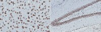
Immunohistochemistry (Paraffin) Analysis: A 1:50-250 dilution from a representative lot detected Lamin A and C in rat cerebral cortex, rat hippocampus, rat cerebellum, and human liver tissue sections.
Biological Information
Immunogen
GST/His-tagged recombinant fragment corresponding to 148 amino acids from the internal region of rat Lamin A/C.
Clone
2F4.1
Concentration
Please refer to lot specific datasheet.
Host
Mouse
Specificity
Clone 2F4.1 detects Lamin A/C in human and rat tissues. It targets an epitope within 148 amino acids from the internal region.
~74/65 kDa observed. Uncharacterized bands may be observed in some lysate(s).
Physicochemical Information
Dimensions
Materials Information
Toxicological Information
Safety Information according to GHS
Safety Information
Product Usage Statements
Quality Assurance
Evaluated by Western Blotting in WI-38 cell lysate.
Western Blotting Analysis: 0.5 µg/mL of this antibody detected Lamin A and C in WI-38 cell lysate.
Usage Statement
Unless otherwise stated in our catalog or other company documentation accompanying the product(s), our products are intended for research use only and are not to be used for any other purpose, which includes but is not limited to, unauthorized commercial uses, in vitro diagnostic uses, ex vivo or in vivo therapeutic uses or any type of consumption or application to humans or animals.
Storage and Shipping Information
Storage Conditions
Stable for 1 year at 2-8°C from date of receipt.
Packaging Information
Material Size
100 μg
Transport Information
Supplemental Information
Specifications
Global Trade Item Number
Bestellnummer
GTIN
MABT545-100UG
04054839507199
Documentation
Anti-Lamin A and C Antibody, clone 2F4.1 Analysenzertifikate