Wenn Sie das Fenster schließen, wird Ihre Konfiguration nicht gespeichert, es sei denn, Sie haben Ihren Artikel in die Bestellung aufgenommen oder zu Ihren Favoriten hinzugefügt.
Klicken Sie auf OK, um das MILLIPLEX® MAP-Tool zu schließen oder auf Abbrechen, um zu Ihrer Auswahl zurückzukehren.
Wählen Sie konfigurierbare Panels & Premixed-Kits - ODER - Kits für die zelluläre Signaltransduktion & MAPmates™
Konfigurieren Sie Ihre MILLIPLEX® MAP-Kits und lassen sich den Preis anzeigen.
Konfigurierbare Panels & Premixed-Kits
Unser breites Angebot enthält Multiplex-Panels, für die Sie die Analyten auswählen können, die am besten für Ihre Anwendung geeignet sind. Unter einem separaten Register können Sie das Premixed-Cytokin-Format oder ein Singleplex-Kit wählen.
Kits für die zelluläre Signaltransduktion & MAPmates™
Wählen Sie gebrauchsfertige Kits zur Erforschung gesamter Signalwege oder Prozesse. Oder konfigurieren Sie Ihre eigenen Kits mit Singleplex MAPmates™.
Die folgenden MAPmates™ sollten nicht zusammen analysiert werden: -MAPmates™, die einen unterschiedlichen Assaypuffer erfordern. -Phosphospezifische und MAPmate™ Gesamtkombinationen wie Gesamt-GSK3β und Gesamt-GSK3β (Ser 9). -PanTyr und locusspezifische MAPmates™, z.B. Phospho-EGF-Rezeptor und Phospho-STAT1 (Tyr701). -Mehr als 1 Phospho-MAPmate™ für ein einziges Target (Akt, STAT3). -GAPDH und β-Tubulin können nicht mit Kits oder MAPmates™, die panTyr enthalten, analysiert werden.
.
Bestellnummer
Bestellinformationen
St./Pkg.
Liste
Dieser Artikel wurde zu Ihren Favoriten hinzugefügt.
Wählen Sie bitte Spezies, Panelart, Kit oder Probenart
Um Ihr MILLIPLEX® MAP-Kit zu konfigurieren, wählen Sie zunächst eine Spezies, eine Panelart und/oder ein Kit.
Custom Premix Selecting "Custom Premix" option means that all of the beads you have chosen will be premixed in manufacturing before the kit is sent to you.
Catalogue Number
Ordering Description
Qty/Pack
List
Dieser Artikel wurde zu Ihren Favoriten hinzugefügt.
Spezies
Panelart
Gewähltes Kit
Menge
Bestellnummer
Bestellinformationen
St./Pkg.
Listenpreis
96-Well Plate
Menge
Bestellnummer
Bestellinformationen
St./Pkg.
Listenpreis
Weitere Reagenzien hinzufügen (MAPmates erfordern die Verwendung eines Puffer- und Detektionskits)
Menge
Bestellnummer
Bestellinformationen
St./Pkg.
Listenpreis
48-602MAG
Buffer Detection Kit for Magnetic Beads
1 Kit
Platzsparende Option Kunden, die mehrere Kits kaufen, können ihre Multiplex-Assaykomponenten in Kunststoffbeuteln anstelle von Packungen erhalten, um eine kompaktere Lagerung zu ermöglichen.
Dieser Artikel wurde zu Ihren Favoriten hinzugefügt.
Das Produkt wurde in Ihre Bestellung aufgenommen
Sie können nun ein weiteres Kit konfigurieren, ein Premixed-Kit wählen, zur Kasse gehen oder das Bestell-Tool schließen.
This mouse monoclonal Anti-PECAM-1 (CD31), clone TLD-3A12, Cat. No. MAB1393-I is tested for use in ELISA, Flow Cytometry, Functional Studies, Immunohistochemistry (Paraffin) and Immunohistochemistry, Immunoprecipitation, and Western Blotting, for the detection of CD31/PECAM-1.
More>>This mouse monoclonal Anti-PECAM-1 (CD31), clone TLD-3A12, Cat. No. MAB1393-I is tested for use in ELISA, Flow Cytometry, Functional Studies, Immunohistochemistry (Paraffin) and Immunohistochemistry, Immunoprecipitation, and Western Blotting, for the detection of CD31/PECAM-1. Less<<
Anti-PECAM-1 (CD31) Antibody, clone TLD-3A12: SDB (Sicherheitsdatenblätter), Analysenzertifikate und Qualitätszertifikate, Dossiers, Broschüren und andere verfügbare Dokumente.
Platelet endothelial cell adhesion molecule (UniProt: Q3SWT0; PECAM-1, CD31) is encoded by the Pecam1 (also known as Pecam) gene (Gene ID: 29583) in rat.PECAM-1 is a single-pass type I membrane protein that contains 6 Ig-like C2-type (immunoglobulin-like) domains. PECAM1 is differentially glycosylated involving both N-linked and O-linked glycosylation sites. It is a cell adhesion molecule that is required for leukocyte trans-endothelial migration (TEM) under most inflammatory conditions. PECAM1 plays a vital role in signaling processes involved in angiogenesis, platelet function, thrombosis, mechanosensing of endothelial cell response to fluid shear stress, and regulation of multiple stages of leukocyte migration through venular walls. PECAM1 is expressed on platelets and leukocytes and is primarily concentrated at the borders between endothelial cells. PECAM1 prevents phagocyte ingestion of closely apposed viable cells by transmitting 'detachment' signals, and changes function on apoptosis, promoting tethering of dying cells to phagocytes. During apoptosis, the inside-out signaling of PECAM1 is disabled that allows the apoptotic cell to accept phagocyte interaction. PECAM1 is phosphorylated on serine and tyrosine residues following cellular activation. In endothelial cells Fyn is shown to mediate mechanical-force induced tyrosine phosphorylation. In response to Fc epsilon RI (FCER1 activation), PECAM1 is phosphorylated on tyrosine residues by FER and FES.
References
Product Information
Format
Purified
Presentation
Purified mouse monoclonal antibody IgG1 in PBS without azide.
This mouse monoclonal Anti-PECAM-1 (CD31), clone TLD-3A12, Cat. No. MAB1393-I is tested for use in ELISA, Flow Cytometry, Functional Studies, Immunohistochemistry (Paraffin) and Immunohistochemistry, Immunoprecipitation, and Western Blotting, for the detection of CD31/PECAM-1.
Key Applications
Immunohistochemistry
Affects Function
Western Blotting
Flow Cytometry
ELISA
Immunohistochemistry (Paraffin)
Application Notes
Immunoprecipitation Analysis: A representative lot detected PECAM-1 (CD31) in Immunoprecipitation applications (Williams, K.C., et. al. (1996). J Neurosci Res. 45(6):747-57).
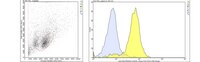
Flow Cytometry Analysis: 1 µg from a representative lot detected PECAM-1 (CD31) in one million rat splenocytes.
Western Blotting Analysis: A representative lot detected PECAM-1 (CD31) in Western Blotting applications (Male, D., et. al. (1995). Immunology. 84(3):453-60).
ELISA Analysis: A representative lot detected PECAM-1 (CD31) in ELISA applications (Male, D., et. al. (1995). Immunology. 84(3):453-60; Williams, K.C., et. al. (1996). J Neurosci Res. 45(6):747-57).
Affects Function: A representative lot of PECAM-1 (CD31) Affected Function (Williams, K.C., et. al. (1996). J Neurosci Res. 45(6):747-57).
Immunohistochemistry Analysis: A representative lot detected PECAM-1 (CD31) in Immunohistochemistry applications (Williams, K.C., et. al. (1996). J Neurosci Res. 45(6):747-57).
Evaluated by Immunohistochemistry in human tonsil.
Immunohistochemistry Analysis: A 1:250 dilution of this antibody detected PECAM-1 (CD31) in human tonsil tissue.
Usage Statement
Unless otherwise stated in our catalog or other company documentation accompanying the product(s), our products are intended for research use only and are not to be used for any other purpose, which includes but is not limited to, unauthorized commercial uses, in vitro diagnostic uses, ex vivo or in vivo therapeutic uses or any type of consumption or application to humans or animals.
Storage and Shipping Information
Storage Conditions
Stable for 1 year at -20°C from date of receipt. Handling Recommendations: Upon receipt and prior to removing the cap, centrifuge the vial and gently mix the solution. Aliquot into microcentrifuge tubes and store at -20°C. Avoid repeated freeze/thaw cycles, which may damage IgG and affect product performance.