Wenn Sie das Fenster schließen, wird Ihre Konfiguration nicht gespeichert, es sei denn, Sie haben Ihren Artikel in die Bestellung aufgenommen oder zu Ihren Favoriten hinzugefügt.
Klicken Sie auf OK, um das MILLIPLEX® MAP-Tool zu schließen oder auf Abbrechen, um zu Ihrer Auswahl zurückzukehren.
Wählen Sie konfigurierbare Panels & Premixed-Kits - ODER - Kits für die zelluläre Signaltransduktion & MAPmates™
Konfigurieren Sie Ihre MILLIPLEX® MAP-Kits und lassen sich den Preis anzeigen.
Konfigurierbare Panels & Premixed-Kits
Unser breites Angebot enthält Multiplex-Panels, für die Sie die Analyten auswählen können, die am besten für Ihre Anwendung geeignet sind. Unter einem separaten Register können Sie das Premixed-Cytokin-Format oder ein Singleplex-Kit wählen.
Kits für die zelluläre Signaltransduktion & MAPmates™
Wählen Sie gebrauchsfertige Kits zur Erforschung gesamter Signalwege oder Prozesse. Oder konfigurieren Sie Ihre eigenen Kits mit Singleplex MAPmates™.
Die folgenden MAPmates™ sollten nicht zusammen analysiert werden: -MAPmates™, die einen unterschiedlichen Assaypuffer erfordern. -Phosphospezifische und MAPmate™ Gesamtkombinationen wie Gesamt-GSK3β und Gesamt-GSK3β (Ser 9). -PanTyr und locusspezifische MAPmates™, z.B. Phospho-EGF-Rezeptor und Phospho-STAT1 (Tyr701). -Mehr als 1 Phospho-MAPmate™ für ein einziges Target (Akt, STAT3). -GAPDH und β-Tubulin können nicht mit Kits oder MAPmates™, die panTyr enthalten, analysiert werden.
.
Bestellnummer
Bestellinformationen
St./Pkg.
Liste
Dieser Artikel wurde zu Ihren Favoriten hinzugefügt.
Wählen Sie bitte Spezies, Panelart, Kit oder Probenart
Um Ihr MILLIPLEX® MAP-Kit zu konfigurieren, wählen Sie zunächst eine Spezies, eine Panelart und/oder ein Kit.
Custom Premix Selecting "Custom Premix" option means that all of the beads you have chosen will be premixed in manufacturing before the kit is sent to you.
Catalogue Number
Ordering Description
Qty/Pack
List
Dieser Artikel wurde zu Ihren Favoriten hinzugefügt.
Spezies
Panelart
Gewähltes Kit
Menge
Bestellnummer
Bestellinformationen
St./Pkg.
Listenpreis
96-Well Plate
Menge
Bestellnummer
Bestellinformationen
St./Pkg.
Listenpreis
Weitere Reagenzien hinzufügen (MAPmates erfordern die Verwendung eines Puffer- und Detektionskits)
Menge
Bestellnummer
Bestellinformationen
St./Pkg.
Listenpreis
48-602MAG
Buffer Detection Kit for Magnetic Beads
1 Kit
Platzsparende Option Kunden, die mehrere Kits kaufen, können ihre Multiplex-Assaykomponenten in Kunststoffbeuteln anstelle von Packungen erhalten, um eine kompaktere Lagerung zu ermöglichen.
Dieser Artikel wurde zu Ihren Favoriten hinzugefügt.
Das Produkt wurde in Ihre Bestellung aufgenommen
Sie können nun ein weiteres Kit konfigurieren, ein Premixed-Kit wählen, zur Kasse gehen oder das Bestell-Tool schließen.
Anti-α-Synuclein, clone 2F12, Cat. No. MABN1817, is a highly specific mouse monoclonal antibody that targets α-synuclein and has been tested in ELISA, Immunocytochemistry, Immunohistochemistry (Paraffin), Immunoprecipitation, and Western Blotting.
More>>Anti-α-Synuclein, clone 2F12, Cat. No. MABN1817, is a highly specific mouse monoclonal antibody that targets α-synuclein and has been tested in ELISA, Immunocytochemistry, Immunohistochemistry (Paraffin), Immunoprecipitation, and Western Blotting. Less<<
Anti-α-Synuclein Antibody, clone 2F12: SDB (Sicherheitsdatenblätter), Analysenzertifikate und Qualitätszertifikate, Dossiers, Broschüren und andere verfügbare Dokumente.
Alpha-synuclein (UniProt P37840; also known as NACP, Non-A beta component of AD amyloid, Non-A4 component of amyloid precursor, Synuclein alpha-140) is encoded by the SNCA (also known as NACP, PARK1, PARK4) gene (Gene ID 6622) in human. Pathological aggregates are common features of many neurodegenerative diseases, such as tau neurofibrillary tangles (NFTs) in Alzheimer’s disease (AD) and frontotemporal degeneration, and α-synuclein (α-syn or αS) Lewy bodies (LBs) in Parkinson’s disease (PD) and dementia with LBs (DLB). Alpha-synuclein is a phospholipid-binding protein concentrated in presynaptic terminals where it promotes SNARE complex formation and modulates synaptic functions. Alpha-synuclein is the major component of pathologic inclusions that characterize PD, DLB, and multiple system atrophy (MSA). Research shows that αS exists not only as unfolded monomers, but in large part also as multimers, principally as ~60 kDa tetramers composed of four N-acetylated αS, that assume α-helical conformation and resist aggregation. PD-causing αS missense mutations are found to shift cellular αS from tetramers/multimers to monomers, indicating that decreased α-helical tetramers and increased unfolded monomers initiate pathogenesis. In addition, both casein kinase-1 (CK-1) and CK-2 can catalyze the phosphorylation of αS on Ser129, and Ser129-phosphorylated αS is found in αS inclusions.
References
Product Information
Format
Purified
Presentation
Purified mouse IgG2b in buffer containing 0.1 M Tris-Glycine (pH 7.4), 150 mM NaCl with 0.05% sodium azide.
Anti-α-Synuclein, clone 2F12, Cat. No. MABN1817, is a highly specific mouse monoclonal antibody that targets α-synuclein and has been tested in ELISA, Immunocytochemistry, Immunohistochemistry (Paraffin), Immunoprecipitation, and Western Blotting.
Key Applications
ELISA
Immunocytochemistry
Immunohistochemistry (Paraffin)
Immunoprecipitation
Western Blotting
Application Notes
Immunohistochemistry Analysis: A 1:1,000 dilution from a representative lot detected α-synuclein in human prostate cancer, cerebral cortex, and kidney tissue sections.
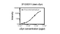
ELISA Analysis: A representative lot (0.4 µL in 30 µL buffer/well for coating) captured recombinant human α-synuclein (0.2-40 ng/mL) in a sandwich ELISA application utilizing clone SOY1 (Cat. No. MABN1818; preconjugated with Sulfo tag) as the detection antibody (Courtesy of Tim Bartels, Ph.D., Brigham and Women's Hospital, Boston, MA, U.S.A.).
Immunocytochemistry Analysis: A 1:1,000 dilution from a representative lot immunostained primary mouse cortical neurons (Courtesy of Tim Bartels, Ph.D., Brigham and Women's Hospital, Boston, MA, U.S.A.).
Immunohistochemistry Analysis: A 1:11,110 dilution from a representative lot immunostained Lewy bodies (LBs) in striatum tissue sections from Parkinson's diseased (PD) human brain (Courtesy of Tim Bartels, Ph.D., Brigham and Women's Hospital, Boston, MA, U.S.A.).
Immunoprecipitation Analysis: 4 µL from a representative lot immunoprecipitated α-synuclein from 50 µg of HEL human erythroleukemia cell lysate (Courtesy of Tim Bartels, Ph.D., Brigham and Women's Hospital, Boston, MA, U.S.A.).
ELISA Analysis: A representative lot captured both endogenous α-synuclein (αS) from human cortical homogenate, as well the exogenously expressed wild type and familial PD (fPD) αS mutants (A30P, E46K, H50Q, G51D, A53T) from sytosolic extracts of transfected M17D human neuroblastoma cells in a sandwich ELISA application utilizing clone SOY1 (Cat. No. MABN1818; preconjugated with Sulfo tag) as the detection antibody (Dettmer, U., et al. (2015). Nat. Commun. 6:7314).
ELISA Analysis: A representative lot captured both pre-aggregated fibrillar recombinant α-synuclein as well as partially purified Lewy bodies (LBs) from a DLB (dementia with LBs) patient with or without prior sample denaturing by boiling with 2% SDS in a sandwich ELISA application utilizing clone SOY1 (Cat. No. MABN1818; preconjugated with Sulfo tag) as the detection antibody (Dettmer, U., et al. (2015). Nat. Commun. 6:7314).
Immunocytochemistry Analysis: A representative lot detected cytosolic localization of endogenous rat α-synuclein (αS) and exogenously overexpressed human αS by fluorescent immunocytochemistry staining of 4% paraformaldehyde-fixed, 0.25% Triton X-100-permeabilized primary rat neurons and transfected M17D human neuroblastoma cells (Dettmer, U., et al. (2015). Nat. Commun. 6:7314).
Western Blotting Analysis: A representative lot detected monomeric α-synuclein (αS) as well as αS multimers (αS60, αS80 and αS100) in extract from disuccinimidyl glutarate (DSG) cross-linked mouse brain bits, human iPSCs (both S A53T mutant and corrected isogenic line) and ESCs (both wild-type and genetically engineered isogenic αS E46K line). A significantly reduced αS60 level was seen with A53T and E46K mutants (Dettmer, U., et al. (2015). Nat. Commun. 6:7314).
Western Blotting Analysis: A representative lot detected monomeric α-synuclein (αS) as well as αS multimers (αS60, αS80 and αS100) in cytosolic extracts from disuccinimidyl glutarate (DSG) cross-linked primary rat neurons, as well as human HEL erythroid leukemia and M17D neuroblastoma cells (Dettmer, U., et al. (2013). J. Biol. Chem. 288(9):6371-6385).
Biological Information
Immunogen
Purified human erythrocyte α-synuclein.
Clone
2F12
Concentration
Please refer to lot specific datasheet.
Host
Mouse
Specificity
Clone 2F12 reacted with both monomeric and aggregated forms of alpha-synuclein of human, mouse, and rat species. Clone 2F12 detected both wild-type alpha-synuclein and fPD mutants (Dettmer, U., et al. (2015). Nat. Commun. 6:7314; Dettmer, U., et al. (2013). J. Biol. Chem. 288(9):6371-6385).
~14.5 kDa observed. 14.46 kDa (human isoform 1; NACP140), 14.49/14.52 kDa (mouse/rat isoform 1) calculated. Uncharacterized bands may be observed in some lysate(s).
Physicochemical Information
Dimensions
Materials Information
Toxicological Information
Safety Information according to GHS
Safety Information
Product Usage Statements
Quality Assurance
Evalulated by Western Blotting in human fetal brain tissue lysate.
Western Blotting Analysis: A 1:1,000 dilution of this antibody detected α-synuclein in 10 µg of human fetal brain tissue lysate.
Usage Statement
Unless otherwise stated in our catalog or other company documentation accompanying the product(s), our products are intended for research use only and are not to be used for any other purpose, which includes but is not limited to, unauthorized commercial uses, in vitro diagnostic uses, ex vivo or in vivo therapeutic uses or any type of consumption or application to humans or animals.