Wenn Sie das Fenster schließen, wird Ihre Konfiguration nicht gespeichert, es sei denn, Sie haben Ihren Artikel in die Bestellung aufgenommen oder zu Ihren Favoriten hinzugefügt.
Klicken Sie auf OK, um das MILLIPLEX® MAP-Tool zu schließen oder auf Abbrechen, um zu Ihrer Auswahl zurückzukehren.
Wählen Sie konfigurierbare Panels & Premixed-Kits - ODER - Kits für die zelluläre Signaltransduktion & MAPmates™
Konfigurieren Sie Ihre MILLIPLEX® MAP-Kits und lassen sich den Preis anzeigen.
Konfigurierbare Panels & Premixed-Kits
Unser breites Angebot enthält Multiplex-Panels, für die Sie die Analyten auswählen können, die am besten für Ihre Anwendung geeignet sind. Unter einem separaten Register können Sie das Premixed-Cytokin-Format oder ein Singleplex-Kit wählen.
Kits für die zelluläre Signaltransduktion & MAPmates™
Wählen Sie gebrauchsfertige Kits zur Erforschung gesamter Signalwege oder Prozesse. Oder konfigurieren Sie Ihre eigenen Kits mit Singleplex MAPmates™.
Die folgenden MAPmates™ sollten nicht zusammen analysiert werden: -MAPmates™, die einen unterschiedlichen Assaypuffer erfordern. -Phosphospezifische und MAPmate™ Gesamtkombinationen wie Gesamt-GSK3β und Gesamt-GSK3β (Ser 9). -PanTyr und locusspezifische MAPmates™, z.B. Phospho-EGF-Rezeptor und Phospho-STAT1 (Tyr701). -Mehr als 1 Phospho-MAPmate™ für ein einziges Target (Akt, STAT3). -GAPDH und β-Tubulin können nicht mit Kits oder MAPmates™, die panTyr enthalten, analysiert werden.
.
Bestellnummer
Bestellinformationen
St./Pkg.
Liste
Dieser Artikel wurde zu Ihren Favoriten hinzugefügt.
Wählen Sie bitte Spezies, Panelart, Kit oder Probenart
Um Ihr MILLIPLEX® MAP-Kit zu konfigurieren, wählen Sie zunächst eine Spezies, eine Panelart und/oder ein Kit.
Custom Premix Selecting "Custom Premix" option means that all of the beads you have chosen will be premixed in manufacturing before the kit is sent to you.
Catalogue Number
Ordering Description
Qty/Pack
List
Dieser Artikel wurde zu Ihren Favoriten hinzugefügt.
Spezies
Panelart
Gewähltes Kit
Menge
Bestellnummer
Bestellinformationen
St./Pkg.
Listenpreis
96-Well Plate
Menge
Bestellnummer
Bestellinformationen
St./Pkg.
Listenpreis
Weitere Reagenzien hinzufügen (MAPmates erfordern die Verwendung eines Puffer- und Detektionskits)
Menge
Bestellnummer
Bestellinformationen
St./Pkg.
Listenpreis
48-602MAG
Buffer Detection Kit for Magnetic Beads
1 Kit
Platzsparende Option Kunden, die mehrere Kits kaufen, können ihre Multiplex-Assaykomponenten in Kunststoffbeuteln anstelle von Packungen erhalten, um eine kompaktere Lagerung zu ermöglichen.
Dieser Artikel wurde zu Ihren Favoriten hinzugefügt.
Das Produkt wurde in Ihre Bestellung aufgenommen
Sie können nun ein weiteres Kit konfigurieren, ein Premixed-Kit wählen, zur Kasse gehen oder das Bestell-Tool schließen.
Milli-Mark ChromaPan Neuronal Marker-ORC is an antibody targeting the ORC protein, validated for use in ICC & IF.
More>>Milli-Mark ChromaPan Neuronal Marker-ORC is an antibody targeting the ORC protein, validated for use in ICC & IF. Less<<
Milli-Mark™ ChromaPan Neuronal Marker-ORC: SDB (Sicherheitsdatenblätter), Analysenzertifikate und Qualitätszertifikate, Dossiers, Broschüren und andere verfügbare Dokumente.
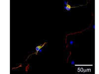
The Milli-Mark™ ChromaPan Neuronal Marker-ORC is a complete multi-species neuronal antibody blend with an open rabbit channel (ORC) that specifically detects axons, dendrites, and cell bodies with different fluorophores. The Multi-species Primary Antibody blend does not have a rabbit IgG; this allows the researcher to colocalize their own rabbit IgG (against their target protein) to the neuronal cytoarchitecture.
Antibodies to neuronal proteins have become critical tools for identifying neurons and discerning morphological characteristics in culture and complex tissue. Neuron-specific antibodies are convenient precision tools useful in revealing cytoarchitecture, but are limited to the protein target distribution within the neuron, which may differ greatly from nucleus to soma to dendrite and axon. To achieve as complete a morphological staining as possible across all parts of neurons, Millipore has developed a multi-species primary antibody blend that reacts against key somatic, dendritic, and axonal proteins distributed across the neuronal architecture that can then be detected by a single multi-species secondary antibody blend containing discrete fluorophores. As a result, different morphological features (somatic, dendritic, and axonal) are illuminated in different colors: The somato-dendritic cytoarchitecture is detected with DyLight® 488 conjugated secondary, and the somato-axonal cytoarchitecture is detected with DyLight® 649 conjugated secondary antibody. The primary antibody blend does not contain any rabbit IgGs. The researcher can add a rabbit IgG against their target protein which can be detected with the secondary antibody blend provided by a cyanine 3 conjugated secondary against Rabbit IgG. This pan-neuronal antibody cocktail has been validated in a variety of cell and tissue cultures, giving researchers a convenient and specific qualitative and quantitative tool for studying neuronal morphology.
References
Product Information
Components
1) 5X Blocking Buffer (Part No. CS204291): 5 ml of an optimized blocking buffer is provided and contains PBS, BSA, serum, trition X-100, and 0.05% azide. Store at 2-8°C.
2) Multi-species Primary Antibody blend (ORC) (Part No. CS204300): 100uL of a proprietary antibody blend with host specific antibodies that reacts against key somatic, nuclear, dendritic and axonal targets. It does not contain rabbit IgG. Presented in PBS containing 0.05% azide. Store at 2-8°C.
3) Multi-species Secondary Antibody blend (Part No. CS204289): 100 µL of a secondary antibody blend containing host specific IgG antibodies against mouse, rabbit, and chicken, conjugated to DyLight 488, cyanine 3, and DyLight 649 dyes respectively. Each individual set has been species absorbed against H, R, B, Gt, Sh, Gp, Eq and/or M, Rb, Ch to minimize background and cross reactivity. Presented in 0.01M Sodium Phosphate, 0.25M NaCl, pH 7.6 with 15 mg/mL BSA, and 0.05% sodium azide. Store at 2-8°C, away from light.
4) DAPI (Part No. CS202687): Vial contains 10 µL of 0.1mg/mL of DAPI. Store at 2-8°C, away from light.
Format
Purified
Control
Cortical neurons
Presentation
Blended polyclonal antibody cocktail in PBS with 0.05% NaN3.
Milli-Mark ChromaPan Neuronal Marker-ORC is an antibody targeting the ORC protein, validated for use in ICC & IF.
Key Applications
Immunocytochemistry
Immunofluorescence
Application Notes
Immunocytochemistry Protocol 1. Culture neurons as desired. When ready, gently wash with PBS and fix cells with 4% paraformaldehyde in PBS, 5-15 min at room temp (RT). 2. Wash with PBS, 3 x 5min each, at RT and block with 1X blocking buffer for 1 hr at RT. Note: The blocking buffer provided is supplied as a 5X concentrate. Dilute the concentrate to 1X strength using Milli-Q™ water. For example, add 20 ml of Milli-Q™ water to 5 ml of 5X blocking buffer. 3. Incubate cells with ChromaPan Multi-species Primary Antibody blend for 2hr at RT (dilute antibody 1:125 to 1:250 in blocking buffer). Optimal working concentrations must be determined by the end user. Optional: Rabbit IgG against target protein can also be added to the ChromaPan primary antibody mixture. Optimal working conditions must be determined by the end user. If rabbit IgG is added skip step 4. 4. Wash with PBS, 3 x 5min each, and incubate cells with rabbit IgG against target protein. Optimal working conditions must be determined by the end user. 5. Wash with PBS, 3 x 5min each, and incubate cells with Multi-species Secondary Antibody blend (1:150 in blocking buffer) for 1hr at RT (validated with Chroma Pan to produce minimal background). Optimal working concentrations must be determined by the end user. Optional: DAPI (1:1000) can also be added to the secondary mixture. If DAPI is added skip step 6. 6. Wash with PBS, 3 x 5min each, and incubate cells with DAPI (1:1000 in blocking buffer) for 5min at RT. Optimal working concentrations must be determined by the end user. 7. Wash with PBS, 3 x 5min each, and cover slip with LIGHT DIAGNOSTICS Mounting Fluid (Catalog No. #5013). View using fluorescent microscope with appropriate filter.
Biological Information
Epitope
Whole Neuron Marker
Host
Mixed Species
Specificity
NS340 is specific to axons, dendrites including spines, and the cell body of neurons.
Species Reactivity
Human
Rat
Mouse
Species Reactivity Note
Rat, Mouse, and Human. Reactivity with other species has not been determined.
Antibody Type
Polyclonal Antibody
Physicochemical Information
Dimensions
Materials Information
Toxicological Information
Safety Information according to GHS
Safety Information
Product Usage Statements
Quality Assurance
Routinely tested on rat E18 cortex primary neurons in Immunocytochemistry 1:150 - 1:100.
Usage Statement
Unless otherwise stated in our catalog or other company documentation accompanying the product(s), our products are intended for research use only and are not to be used for any other purpose, which includes but is not limited to, unauthorized commercial uses, in vitro diagnostic uses, ex vivo or in vivo therapeutic uses or any type of consumption or application to humans or animals.
Storage and Shipping Information
Storage Conditions
Maintain at 2-8°C for up to 6 months from date of receipt.