Si cierra, no se guardará su personalización salvo que haya añadido el artículo a su carrito de la compra o a favoritos.
Pulse OK para cerrar la herramienta MILLIPLEX® MAP o Cancelar para volver a su selección.
Elija paneles personalizables y kits premezclos - O - MAPmates™ de señalización celular
Diseñe y calcule el precio de sus kits MILLIPLEX® MAP.
Paneles personalizados y kits premezclados
Nuestra amplia cartera de productos consta de paneles multiplex que le permiten elegir, dentro del panel, los analitos que mejor se ajustan a sus requisitos. En una pestaña distinta puede elegir el formato de citocina premezclada o un kit single plex.
Kits de señalización celular y MAPmates™
Elija los kits preparados para poder explorar las vías o los procesos enteros. O diseñe sus propios kits eligiendo single plex MAPmates™ según las directrices proporcionadas.
No deben combinarse los siguientes MAPmates™: -MAPmates™ que requieren un tampón de ensayo diferente. -Pares MAPmate™ fosfoespecíficos y totales, por ejemplo, GSK3β y GSK3β (Ser 9). -MAPmates™ con panTyr y específicos de sitio; por ejemplo, receptor del fosfo-EGF y fosfo-STAT1 (Tyr701). -Más de 1 fosfo-MAPmate™ para una sola diana (Akt, STAT3). -La GAPDH y la β-tubulina no pueden combinarse con kits o MAPmates™ que contengan panTyr.
.
Número de referencia
Descripción para pedidos
Cant./Env.
Lista
Este artículo se ha añadido a favoritos.
Seleccione una especie, un tipo de panel, un kit o un tipo de muestra
Para empezar a diseñar su kit MILLIPLEX® MAP, seleccione una especie, un tipo de panel o un kit de interés.
Custom Premix Selecting "Custom Premix" option means that all of the beads you have chosen will be premixed in manufacturing before the kit is sent to you.
Catalogue Number
Ordering Description
Qty/Pack
List
Este artículo se ha añadido a favoritos.
Especie
Tipo de panel
Kit seleccionado
Cant.
Número de referencia
Descripción para pedidos
Cant./Env.
Precio de catálogo
96-Well Plate
Cant.
Número de referencia
Descripción para pedidos
Cant./Env.
Precio de catálogo
Añadir más reactivos (Se necesita tampón y un kit de detección para usar con MAPmates)
Cant.
Número de referencia
Descripción para pedidos
Cant./Env.
Precio de catálogo
48-602MAG
Buffer Detection Kit for Magnetic Beads
1 Kit
Opción para ahorrar espacio Los clientes que adquieran múltiples kits pueden optar por ahorrar espacio de almacenamiento retirando el embalaje del kit y recibiendo los componentes de sus ensayos multiplex en bolsas de plástico para un almacenamiento más compacto.
Este artículo se ha añadido a favoritos.
El producto se ha añadido a su carrito
Ahora puede personalizar otro kit, elegir un kit premezclado, tramitarlo o cerrar la herramienta de pedidos.
The goal of drug discovery is to find well-qualified candidate drugs expected to provide therapeutic benefit. Use of relevant and information-rich assays can rapidly eliminate candidates that either do not affect the appropriate response or cause some unforeseen lethality, and can save drug companies time and money that is often wasted pursuing clinical trials with flawed drug candidates. Many of the Amnis® imaging flow cytometry's applications are ideally suited for characterizing drug candidates, including cell signaling, cell-cell interactions, chemokine-induced shape change, cell death, co-localization, and phagocytosis, to name a few. Furthermore, these applications work with primary cells such as whole blood leukocytes and can often be multiplexed.
Co-Localization of an Antibody-Drug Conjugate to Endosomes or Lysosomes
In this experiment we use the capabilities of the Imagestream®x system to measure the transit of a fluorescently labeled antibody-drug conjugate (ADC) through the cellular endocytic pathway. The high spatial resolution afforded by the Imagestream®x system allowed accurate measurement of ADC co-localization to endosomes and lysosomes. See the application note for more details.
Barcoding
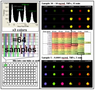
Screening drug candidates requires the rapid analysis of large numbers of samples. Multiplexing samples on the Imagestream®x by mixing dozens of samples from a time course or dose response titration into one tube dramatically increases the sample throughput of the system. This is achieved through fluorescent cell barcoding with subsequent identification of each sample based on a pattern of fluorescent dye and staining intensity unique to each treatment/time point. Automated location of populations based on fluorescence intensity and color allows deconvolution of the barcoded samples after acquisition.
Here is an example of measuring the movement of NF-κB from the cytoplasm to the nucleus in response to varying doses and times of exposure to TNF-α. By labeling 64 separate samples with different combinations of 3 dyes (AlexaFluor 405, 488, and 660) at 4 intensity levels (43 = 64) it was possible to run one tube of the combined samples on the Imagestream®x and acquire a single file. After acquisition of over 600,000 images in 10-15 minutes, the color and intensity combinations of the samples were deconvoluted for further quantitative image analysis. Mean Similarity scores are reported in the spreadsheet and a heatmap has been applied. See the cell signaling application for more information about the translocation assay. The combination of high speed image acquisition and automated quantitative image analysis provided by the Imagestream®x system offers a major advancement in the field of drug discovery.