


CP64 Sigma-AldrichAnti-Actin (Ab-3) Mouse mAb (HHF-35)

Recommended Products
Overview
| Replacement Information |
|---|
Key Spec Table
| Host |
|---|
| M |
| Description | |
|---|---|
| Overview | This product has been discontinued. We are offering Anti-Actin Antibody, clone C4 (Cat. No. MAB1501) as a possible alternative. Please read the alternative product documentation carefully and contact technical service if you need additional information. |
| Catalogue Number | CP64 |
| Brand Family | Calbiochem® |
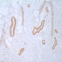
| Application Data |  Detection of actin by staining paraffin sections. Sample: Skeletal muscle. Primary antibody: Anti-Actin (Ab-3) Mouse mAb (HHF-35) (Cat. No. CP64) Detection: colorimetric (DAB). |
| References | |
|---|---|
| References | Schmidt, R.A., et al. 1988. Am. J. Pathol. 131, 19. Tsukada, T., et al. 1987. Am. J. Pathol. 127, 389. Gown, A.M., et al. 1986. Am. J. Pathol. 125, 191. |
| Product Information | |
|---|---|
| Form | Liquid |
| Formulation | Ascites diluted in proprietary buffer. |
| Negative control | Normal mouse serum |
| Positive control | Skeletal muscle tissue |
| Preservative | ≤0.2% sodium azide |
| Biological Information | |
|---|---|
| Immunogen | an SDS extract from human myocardium |
| Immunogen | Human |
| Clone | HHF-35 |
| Host | Mouse |
| Isotype | IgG |
| Physicochemical Information |
|---|
| Dimensions |
|---|
| Materials Information |
|---|
| Toxicological Information |
|---|
| Safety Information according to GHS |
|---|
| Safety Information |
|---|
| Product Usage Statements |
|---|
| Storage and Shipping Information | |
|---|---|
| Ship Code | Blue Ice Only |
| Toxicity | Highly Toxic |
| Storage | +2°C to +8°C |
| Do not freeze | Yes |
| Packaging Information |
|---|
| Transport Information |
|---|
| Supplemental Information |
|---|
| Specifications |
|---|
| Global Trade Item Number | |
|---|---|
| Catalogue Number | GTIN |
| CP64 | 0 |
Documentation
Anti-Actin (Ab-3) Mouse mAb (HHF-35) SDS
| Title |
|---|
References
| Reference overview |
|---|
| Schmidt, R.A., et al. 1988. Am. J. Pathol. 131, 19. Tsukada, T., et al. 1987. Am. J. Pathol. 127, 389. Gown, A.M., et al. 1986. Am. J. Pathol. 125, 191. |
Brochure
| Title |
|---|
| Antibody Sourcebookル Sixth Edition |
| Stem Cell Resource Brochure GBP |