Mixed Electrical-Chemical Synapses in Adult Rat Hippocampus are Primarily Glutamatergic and Coupled by Connexin-36.
Hamzei-Sichani, F; Davidson, KG; Yasumura, T; Janssen, WG; Wearne, SL; Hof, PR; Traub, RD; Gutiérrez, R; Ottersen, OP; Rash, JE
Frontiers in neuroanatomy
6
13
2012
Show Abstract
Dendrodendritic electrical signaling via gap junctions is now an accepted feature of neuronal communication in mammalian brain, whereas axodendritic and axosomatic gap junctions have rarely been described. We present ultrastructural, immunocytochemical, and dye-coupling evidence for "mixed" (electrical/chemical) synapses on both principal cells and interneurons in adult rat hippocampus. Thin-section electron microscopic images of small gap junction-like appositions were found at mossy fiber (MF) terminals on thorny excrescences of CA3 pyramidal neurons (CA3pyr), apparently forming glutamatergic mixed synapses. Lucifer Yellow injected into weakly fixed CA3pyr was detected in MF axons that contacted four injected CA3pyr, supporting gap junction-mediated coupling between those two types of principal cells. Freeze-fracture replica immunogold labeling revealed diverse sizes and morphologies of connexin-36-containing gap junctions throughout hippocampus. Of 20 immunogold-labeled gap junctions, seven were large (328-1140 connexons), three of which were consistent with electrical synapses between interneurons; but nine were at axon terminal synapses, three of which were immediately adjacent to distinctive glutamate receptor-containing postsynaptic densities, forming mixed glutamatergic synapses. Four others were adjacent to small clusters of immunogold-labeled 10-nm E-face intramembrane particles, apparently representing extrasynaptic glutamate receptor particles. Gap junctions also were on spines in stratum lucidum, stratum oriens, dentate gyrus, and hilus, on both interneurons and unidentified neurons. In addition, one putative GABAergic mixed synapse was found in thin-section images of a CA3pyr, but none were found by immunogold labeling, suggesting the rarity of GABAergic mixed synapses. Cx36-containing gap junctions throughout hippocampus suggest the possibility of reciprocal modulation of electrical and chemical signals in diverse hippocampal neurons. | 22615687
 |
Tissue-specific cross-reactivity of connexin32 antibodies: problems and solutions unique to the central nervous system.
Fowler, Stephanie L, et al.
Cell Commun. Adhes., 16: 117-30 (2009)
2009
Show Abstract
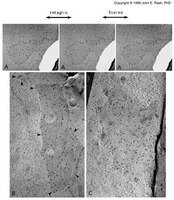
Gap junction proteins are a highly homologous family of 21 connexins. Here, the authors describe a tissue-specific technical artifact complicating analysis of connexin32 protein expression in the central nervous system. The authors show that in brain, but not liver, eight commonly employed antibodies exhibit a higher affinity for a cross-reactive protein that masks the detection of connexin32. Cross-reactivity is evident in Western blot analyses when proteins are subjected to reducing/denaturing conditions but not immunoprecipitation or immunofluorescent applications. Through bioinformatic analyses, tested by sucrose gradient fractionation and immunoblotting of lysates from connexin null-mutant mice, the authors show that the cross-reactive protein is not found in the same cellular compartments as connexin32 and is likely not a member of the connexin family. These findings are presented with the intent of helping to reduce the amount of time laboratories currently expend in validating changes in connexin32 expression in the central nervous system. | 19845480
|
Identification of connexin36 in gap junctions between neurons in rodent locus coeruleus.
Rash, JE; Olson, CO; Davidson, KG; Yasumura, T; Kamasawa, N; Nagy, JI
Neuroscience
147
938-56
2007
Show Abstract
Locus coeruleus neurons are strongly coupled during early postnatal development, and it has been proposed that these neurons are linked by extraordinarily abundant gap junctions consisting of connexin32 (Cx32) and connexin26 (Cx26), and that those same connexins abundantly link neurons to astrocytes. Based on the controversial nature of those claims, immunofluorescence imaging and freeze-fracture replica immunogold labeling were used to re-investigate the abundance and connexin composition of neuronal and glial gap junctions in developing and adult rat and mouse locus coeruleus. In early postnatal development, connexin36 (Cx36) and connexin43 (Cx43) immunofluorescent puncta were densely distributed in the locus coeruleus, whereas Cx32 and Cx26 were not detected. By freeze-fracture replica immunogold labeling, Cx36 was found in ultrastructurally-defined neuronal gap junctions, whereas Cx32 and Cx26 were not detected in neurons and only rarely detected in glia. In 28-day postnatal (adult) rat locus coeruleus, immunofluorescence labeling for Cx26 was always co-localized with the glial gap junction marker Cx43; Cx32 was associated with the oligodendrocyte marker 2',3'-cyclic nucleotide 3'-phosphodiesterase (CNPase); and Cx36 was never co-localized with Cx26, Cx32 or Cx43. Ultrastructurally, Cx36 was localized to gap junctions between neurons, whereas Cx32 was detected only in oligodendrocyte gap junctions; and Cx26 was found only rarely in astrocyte junctions but abundantly in pia mater. Thus, in developing and adult locus coeruleus, neuronal gap junctions contain Cx36 but do not contain detectable Cx32 or Cx26, suggesting that the locus coeruleus has the same cell-type specificity of connexin expression as observed ultrastructurally in other regions of the CNS. Moreover, in both developing and adult locus coeruleus, no evidence was found for gap junctions or connexins linking neurons with astrocytes or oligodendrocytes, indicating that neurons in this nucleus are not linked to the pan-glial syncytium by Cx32- or Cx26-containing gap junctions or by abundant free connexons composed of those connexins. | 17601673
|
Connexin36 vs. connexin32, miniature neuronal gap junctions, and limited electrotonic coupling in rodent suprachiasmatic nucleus.
J E Rash,C O Olson,W A Pouliot,K G V Davidson,T Yasumura,C S Furman,S Royer,N Kamasawa,J I Nagy,F E Dudek
Neuroscience
149
2007
Show Abstract
Suprachiasmatic nucleus (SCN) neurons generate circadian rhythms, and these neurons normally exhibit loosely-synchronized action potentials. Although electrotonic coupling has long been proposed to mediate this neuronal synchrony, ultrastructural studies have failed to detect gap junctions between SCN neurons. Nevertheless, it has been proposed that neuronal gap junctions exist in the SCN; that they consist of connexin32 or, alternatively, connexin36; and that connexin36 knockout eliminates neuronal coupling between SCN neurons and disrupts circadian rhythms. We used confocal immunofluorescence microscopy and freeze-fracture replica immunogold labeling to examine the distributions of connexin30, connexin32, connexin36, and connexin43 in rat and mouse SCN and used whole-cell recordings to re-assess electrotonic and tracer coupling. Connexin32-immunofluorescent puncta were essentially absent in SCN but connexin36 was relatively abundant. Fifteen neuronal gap junctions were identified ultrastructurally, all of which contained connexin36 but not connexin32, whereas nearby oligodendrocyte gap junctions contained connexin32. In adult SCN, one neuronal gap junction was >600 connexons, whereas 75% were smaller than 50 connexons, which may be below the limit of detectability by fluorescence microscopy and thin-section electron microscopy. Whole-cell recordings in hypothalamic slices revealed tracer coupling with neurobiotin in <5% of SCN neurons, and paired recordings (>40 pairs) did not reveal obvious electrotonic coupling or synchronized action potentials, consistent with few neurons possessing large gap junctions. However, most neurons had partial spikes or spikelets (often <1 mV), which remained after QX-314 [N-(2,6-dimethylphenylcarbamoylmethyl)triethylammonium bromide] had blocked sodium-mediated action potentials within the recorded neuron, consistent with spikelet transmission via small gap junctions. Thus, a few miniature gap junctions on most SCN neurons appear to mediate weak electrotonic coupling between limited numbers of neuron pairs, thus accounting for frequent detection of partial spikes and hypothetically providing the basis for loose electrical or metabolic synchronization of electrical activity commonly observed in SCN neuronal populations during circadian rhythms. Full Text Article | 17904757
|
Intercellular communication via gap junctions in activated rat hepatic stellate cells.
Richard Fischer, Roland Reinehr, Thuy Phung Lu, Alexandra Schönicke, Ulrich Warskulat, Hans Peter Dienes, Dieter Häussinger
Gastroenterology
128
433-48
2005
Show Abstract
BACKGROUND AND AIMS: Gap junctional communication was studied in quiescent and activated hepatic stellate cells. METHODS: Connexin expression and intercellular dye transfer were studied in rat hepatic stellate cells in culture and in vivo. RESULTS: Protein expression of connexin 43 was up-regulated in activated hepatic stellate cells in vivo and in vitro and was mainly localized on the cell surface, whereas connexin 26 was found intracellularly. In contrast to hepatocytes, hepatic stellate cells do not express connexin 32. Confluent hepatic stellate cells in culture communicate via gap junctions, resulting in lucifer yellow transfer and propagation of intracellular calcium signals. Phorbol ester induces a protein kinase C-dependent hyperphosphorylation and degradation of connexin 43 and inhibits intercellular communication on a short-term time scale. At the long-term level, vitamin D(3) , lipopolysaccharide, thyroid hormone T(3), dexamethasone, platelet-derived growth factor, endothelin 1, and interleukin 1beta up-regulate connexin 43 protein and messenger RNA expression and enhance intercellular communication. Slight down-regulation of connexin 43 is observed in response to vitamin A. Connexin 43 induction by endothelin 1 is inhibited by both endothelin A and endothelin B receptor antagonists. In coculture systems, hepatic stellate cells communicate with each other, which is suggestive of a syncytial organization, but no communication was found between hepatic stellate cells and other liver cell types. As shown by immunohistochemistry and electron microscopy, gap junctions are formed between activated hepatic stellate cells in vivo. CONCLUSIONS: Gap junctional communication occurs between hepatic stellate cells, is enhanced after activation, and underlies complex regulation by cytokines, hormones, and vitamins. | 15685554
|
High-resolution proteomic mapping in the vertebrate central nervous system: close proximity of connexin35 to NMDA glutamate receptor clusters and co-localization of connexin36 with immunoreactivity for zonula occludens protein-1 (ZO-1).
J E Rash, A Pereda, N Kamasawa, C S Furman, T Yasumura, K G V Davidson, F E Dudek, C Olson, X Li, J I Nagy
Journal of neurocytology
33
131-51
2004
Show Abstract
Combined confocal microscopy and freeze-fracture replica immunogold labeling (FRIL) were used to examine the connexin identity at electrical synapses in goldfish brain and rat retina, and to test for co-localization vs. close proximity of connexins to other functionally interacting proteins in synapses of goldfish and mouse brain and rat retina. In goldfish brain, confocal microscopy revealed immunofluorescence for connexin35 (Cx35) and NMDA-R1 (NR1) glutamate receptor protein in Mauthner Cell/Club Ending synapses. By FRIL double labeling, NR1 glutamate receptors were found in clusters of intramembrane particles in the postsynaptic membrane extraplasmic leaflets, and these distinctive postsynaptic densities were in close proximity (0.1-0.3 microm) to neuronal gap junctions labeled for Cx35, which is the fish ortholog of connexin36 (Cx36) found at neuronal gap junctions in mammals. Immunogold labeling for Cx36 in adult rat retina revealed abundant gap junctions, including several previously unrecognized morphological types. As in goldfish hindbrain, immunogold double labeling revealed NR1-containing postsynaptic densities localized near Cx36-labeled gap junction in rat inferior olive. Confocal immunofluorescence microscopy revealed widespread co-localization of Cx36 and ZO-1, particularly in the reticular thalamic nucleus and amygdala of mouse brain. By FRIL, ZO-1 immunoreactivity was co-localized with Cx36 at individual gap junction plaques in rat retinal neurons. As cytoplasmic accessory proteins, ZO-1 and possibly related members of the membrane-associated guanylate kinase (MAGUK) family represent scaffolding proteins that may bind to and regulate the activity of many neuronal gap junctions. These data document the power of combining immunofluorescence confocal microscopy with FRIL ultrastructural imaging and immunogold labeling to determine the relative proximities of proteins that are involved in short- vs. intermediate-range molecular interactions in the complex membrane appositions at synapses between neurons. Full Text Article | 15173637
|
Functional coupling between neurons and glia.
V Alvarez-Maubecin, F Garcia-Hernandez, J T Williams, E J Van Bockstaele
The Journal of neuroscience : the official journal of the Society for Neuroscience
20
4091-8
2000
Show Abstract
Neuronal-glial interactions play an important role in information processing in the CNS. Previous studies have indicated that electrotonic coupling between locus ceruleus (LC) neurons is involved in synchronizing the spontaneous activity. The results of the present study extend the functional electrotonic coupling to interactions between neurons and glia. Spontaneous oscillations in the membrane potential were observed in a subset of glia. These oscillations were synchronous with the firing of neurons, insensitive to transmitter receptor antagonists and disrupted by carbenoxolone, a gap junction blocker. Hyperpolarization of neurons with [Met] (5)enkephalin blocked the oscillations in glia. Selective depolarization of glia with a glutamate transporter substrate (l-alpha-aminoadipic acid) increased the neuronal firing rate, suggesting that changes in the membrane potential of glia can modulate neuronal excitability through heterocellular coupling. Dye-coupling experiments further confirmed that small molecules could be transferred through gap junctions between these distinct cell types. No dye transfer was observed between neurons and oligodendrocytes or between astrocytes and oligodendrocytes, suggesting that the junctional communication was specific for astrocytes and neurons. Finally, immunoelectron microscopy studies established that connexins, the proteins that form gap junctions, were present on portions of the plasmalemma, bridging the cytoplasm of neurons and glia in LC. This heterocellular coupling extends the mechanisms by which glia participate in the network properties of the LC in which the degree of coupling is thought to influence cognitive performance. | 10818144
|
Endometrial connexin expression in the mare and pig: evidence for the suppression of cell-cell communication in uterine luminal epithelium.
W E Day, J A Bowen, R Barhoumi, F W Bazer, R C Burghardt
The Anatomical record
251
277-85
1998
Show Abstract
This investigation examines the relationship between implantation strategy and gap junction protein expression in uterine endometrium. The pattern of gap junction and connexin protein expression was analyzed in porcine and equine endometrium from cycling and pregnant animals using electron microscopy and immunocytochemistry. Functional analysis of cell-cell communication was also monitored by laser cytometry in primary cultures of endometrial epithelial cells. Gap junctions were detected in endometrial stroma of cycling and pregnant animals, which was correlated with immunoreactive Cx43 within stromal fibroblasts and vascular elements. No Cx26, Cx32, or Cx43 immunostaining was detected in luminal endometrial epithelium in either the mare or the pig at any stage of the estrous cycle or pregnancy. In contrast, endometrial glands of the mare exhibited a spatiotemporal pattern of Cx43 expression in the apicolateral plasma membrane which, when present, colocalized with the tight junction-associated protein, ZO-1. Uterine glandular Cx43 expression in mares was present from day 3 postovulation through day 14 of diestrus and until day 23 of pregnancy, whereas Cx43 was absent within uterine glands during seasonal anestrus, estrus, and after day 30 of pregnancy. Primary cultures of equine endometrial epithelial cells expressed both immunoreactive Cx43 and significant gap junction-mediated intercellular communication (GJIC) which was rapidly upregulated by 1.0 mM 8-bromo-cAMP or blocked with 1.0 mM octanol. No GJIC or connexin protein was detected in cultured porcine epithelial cells despite incubation with a variety of agents, including 8-bromo-cAMP, steroid hormones, retinoic acid, and/or prolactin. Junctional communication in endometrial epithelium of domestic farm animals is different than that reported for species exhibiting invasive implantation. The absence of GJIC in uterine luminal epithelium of the gilt and mare may be involved in limiting trophoblast invasiveness. | 9669753
|
Topological distribution of two connexin32 antigenic sites in intact and split rodent hepatocyte gap junctions.
Goodenough, D A, et al.
J. Cell Biol., 107: 1817-24 (1988)
1988
Show Abstract
The membrane topology of connexin32, a principal polypeptide of gap junctions in diverse cell types, has been studied in rat and mouse hepatocyte gap junctions using site-specific antisera raised against synthetic oligopeptides corresponding to amino acid sequences deduced from cDNA clones. Based on published hydropathicity maps and identified protease-sensitive cleavage sites, oligopeptides were synthesized corresponding to two hydrophilic domains of connexin32, one predicted to face the cytoplasm, the other predicted to be directed extracellularly. Antisera were raised to keyhole limpet hemocyanin conjugates of the oligopeptides and used to map the distribution of their antigens using indirect immunocytochemistry on isolated gap junctions. The results directly demonstrated the cytoplasmic orientation of an antigen contained within amino acids 98-124 of the connexin32 sequence. The extracellular space in intact, isolated gap junctions is too small to permit binding of antibody molecules, necessitating the experimental separation of the junctional membranes to expose their extracellular surfaces using a urea/alkali procedure. While an antigen contained within amino acids 164-189 was visualized on the extracellular surfaces of some of the separated junctional membranes, variability in the observations and in the splitting procedure left ambiguities concerning the biological relevance of the observations after the denaturing conditions necessary to separate the junctional membranes. Using a different approach, however, the antigen could be exposed in intact liver using a hypertonic disaccharide junction-splitting procedure. The period of time of antigen exposure at the cell surface appears to peak at 30 s and disappear by 2-4 min. Taken together, these data demonstrate the extracellular orientation of an antigen contained within amino acids 164-189, which may be involved in cell-cell interaction within the gap junction. | 2460469
|