


AP1021 Sigma-AldrichAnti-Cytokeratin 18 Rabbit pAb

Recommended Products
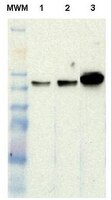
Overview
| Replacement Information |
|---|
Key Spec Table
| Host |
|---|
| Rb |

| Product Information | |
|---|---|
| Form | Liquid |
| Formulation | In PBS. |
| Positive control | MCF-7 cells |
| Preservative | ≤0.1% sodium azide |
| Physicochemical Information |
|---|
| Dimensions |
|---|
| Materials Information |
|---|
| Toxicological Information |
|---|
| Safety Information according to GHS |
|---|
| Safety Information |
|---|
| Product Usage Statements |
|---|
| Packaging Information |
|---|
| Transport Information |
|---|
| Supplemental Information |
|---|
| Specifications |
|---|
| Global Trade Item Number | |
|---|---|
| Catalogue Number | GTIN |
| AP1021 | 0 |
Documentation
Anti-Cytokeratin 18 Rabbit pAb Certificates of Analysis
| Title | Lot Number |
|---|---|
| AP1021 |
References
| Reference overview |
|---|
| Ditzel, H., et al. 2002. J. Biol. Chem. 277, 21712-21722. Caulin, C., et al. 1997. J. Cell Biol. 138, 1379-1394. Oshima, R.G., et al. 1996. Cancer Metastasis Rev. 15, 445-471. Fuchs, E., and Weber, K. 1994. Annu. Rev. Biochem. 63, 345-382. |