


ST1630 Sigma-AldrichAnti-FGF8 Mouse mAb (2A10)


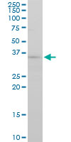
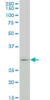
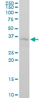
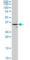
Anti-FGF8, mouse monoclonal, clone 2A10, recognizes the ~30-35 kDa FGF8 protein in PC-12, Jurkat, HepG2, NIH3T3, and Raw 264.7 cells. It is validated for use in ELISA and Western blotting.
More>> Anti-FGF8, mouse monoclonal, clone 2A10, recognizes the ~30-35 kDa FGF8 protein in PC-12, Jurkat, HepG2, NIH3T3, and Raw 264.7 cells. It is validated for use in ELISA and Western blotting. Less<<Anti-FGF8 Mouse mAb (2A10) MSDS (material safety data sheet) or SDS, CoA and CoQ, dossiers, brochures and other available documents.
Synonyms: Anti-Fibroblast Growth Factor 8
Recommended Products
Overview
| Replacement Information |
|---|
Key Spec Table
| Species Reactivity | Host | Antibody Type |
|---|---|---|
| H, M, R | M | Monoclonal Antibody |
Pricing & Availability
| Catalogue Number | Availability | Packaging | Qty/Pack | Price | Quantity | |
|---|---|---|---|---|---|---|
| ST1630-100UG |
|
100 μg |
|
— |
| References |
|---|
| Product Information | |
|---|---|
| Form | Liquid |
| Formulation | In PBS, pH 7.2. |
| Positive control | PC-12 cells, Jurkat cells, HepG2 cells, NIH3T3 cells, Raw 264.7 cells |
| Preservative | None |
| Quality Level | MQ100 |
| Physicochemical Information |
|---|
| Dimensions |
|---|
| Materials Information |
|---|
| Toxicological Information |
|---|
| Safety Information according to GHS |
|---|
| Safety Information |
|---|
| Product Usage Statements |
|---|
| Packaging Information |
|---|
| Transport Information |
|---|
| Supplemental Information |
|---|
| Specifications |
|---|
| Global Trade Item Number | |
|---|---|
| Catalogue Number | GTIN |
| ST1630-100UG | 04055977223736 |
Documentation
Anti-FGF8 Mouse mAb (2A10) SDS
| Title |
|---|