コンテンツへスキップ
Merck
ホーム細胞計数・セルヘルス分析細胞生存率と細胞増殖のMTTアッセイプロトコル

細胞生存率と細胞増殖のMTTアッセイプロトコル

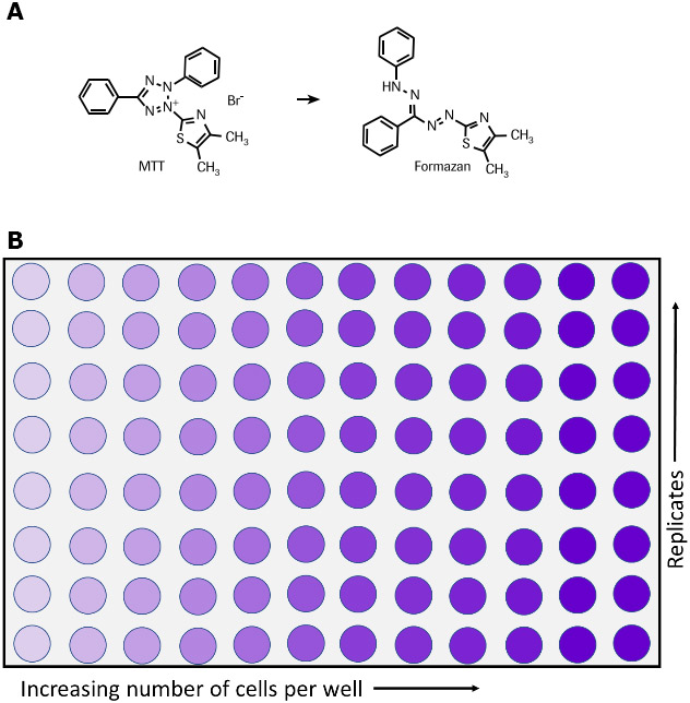
MTTアッセイは、細胞生存率、細胞増殖、および細胞毒性の指標として細胞の代謝活性を測定するために使用されます。この比色分析法では、黄色テトラゾリウム塩(3-(4, 5-ジメチル-チアゾール-2-イル)-2, 5-ジフェニルテトラゾリウムブロマイド、略してMTT)を紫色ホルマザン結晶に還元する細胞の代謝活性を測定します(図1)6、7、35。生細胞には、MTTをホルマザンに還元するNAD(P)H依存性酸化還元酵素が含まれています36。不溶性ホルマザン結晶は可溶化溶液を用いて溶解され、得られた着色溶液は、マルチウェル分光光度計を使用して500〜600ナノメートルでの吸光度を測定することによって定量化されます。溶液の色が濃いほど、代謝活性を持つ生細胞の数が多くなります。

MTTを使用したこの非放射性比色分析システムは、T Mosmann他1によって最初に提唱された後、他の研究者たちによって改良されました2-6。細胞増殖キットI(MTT)は、すぐに使用できる試薬を含む最適化されたMTTアッセイキットであり、洗浄ステップや追加の試薬を必要としません。多数のサンプルを迅速かつ効率的に処理できる定量アッセイです。細胞増殖キットI(MTT)は、次のような複数の用途に使用できます。

  • 細胞増殖と細胞生存率の定量化1、3、5-7
  • 成長因子、サイトカインおよび栄養素に応答した細胞増殖の測定1-3、6、8-12(図3を参照)
  • 細胞毒性の測定。たとえば、腫瘍壊死因子TNF-aまたはTNF-bの影響13、14(図2を参照)あるいはマクロファージ誘導性細胞死の定量化15、16、細胞毒性17-34または阻害抗体などの成長阻害剤の評価など
  • 細胞の活性化の研究4
ホルマザン塩および96ウェルプレートへのMTTの代謝

図1.化学反応(A)および96ウェルプレート(B)で示す、生細胞によるMTTのホルマザン塩への代謝

細胞増殖キットI(MTT)の内容(製品番号11465007001

MTT試薬

  • 調製済み、非滅菌
  • 5 ml MTT(3- [4,5-ジメチルチアゾール-2-イル] -2,5-ジフェニルテトラゾリウムブロミド)(1X)を含むバイアル5個、リン酸緩衝生理食塩水(PBS、806552)中5 mg/ml

可溶化溶液

  • 1X、調製済み
  • 90 mL入りボトル3本。

細胞毒性測定用のアッセイプロトコル

その他の必要な試薬:

  • 培地:10%非働化済 FCS(ウシ胎児血清、12106C)、2 mMグルタミン(G6392)、1 μg/mLアクチノマイシンC1(アクチノマイシンD、A9415)含有RPMI 1640 (R0883)など
  • 抗生物質を使用する場合、ペニシリン/ストレプトマイシンまたはゲンタマイシン含有培地を追加補充する
  • ヒト腫瘍壊死因子-α(hTNF-α)(10 µg/mL)、滅菌済(T6674

プロトコル:

WEHI-164細胞(マウス線維肉腫、87022501)に対するヒト腫瘍壊死因子-α(hTNF-α、T6674)の細胞毒性効果を測定するため(図2参照)。

  1. 1 × 10⁶細胞/mlの濃度のWEHI-164細胞を、1 µg/mLアクチノマイシンC1を含んだ培地で37 ℃および5〜6.5% CO2の条件下で3時間プレインキュベートする
  2. マイクロプレート(組織培養グレード、96ウェル、平底)に、細胞を5 × 10⁴cells/ウェル、1 μg/mLアクチノマイシンC1・さまざまな量のhTNF-α(最終濃度、例えば0.001~0.5 ng/mL)を含有する培養培地100 μLの濃度で播種する
  3. 細胞培養を+37 ℃および5〜6.5% CO2の条件下で24時間インキュベートする
  4. インキュベーション期間の後で、10 µLのMTT標識試薬(最終濃度は0.5 mg/ml)を各ウェルに加える
  5. マイクロプレートを高湿度環境下(+37 ℃、5〜6.5% CO2など)で4時間インキュベートする
  6. 100 µLの可溶化溶液を各ウェルに加える
  7. 高湿度環境下(+37 ℃、5〜6.5% CO2など)のインキュベータ内でプレートを一晩放置する
  8. 紫色ホルマザン結晶が完全に可溶化されていることを確認し、マイクロプレート(ELISA)リーダーを使用してサンプルの吸光度を測定する。ホルマザン生成物の吸光度を測定する波長は、550〜600 nmである(使用するELISAリーダーに搭載されたフィルターによる)。リファレンス波長は650 nm以上となる。
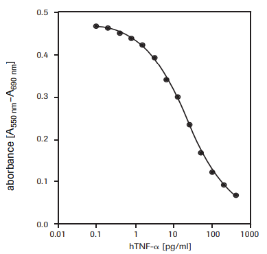
細胞毒性活性の測定

図2.WEHI-164細胞(マウス線維肉腫)に対する組換え型ヒトTNF-α(hTNF-α)の細胞毒性活性をMTTアッセイを使用して測定

細胞増殖測定用のアッセイプロトコル

その他の必要な試薬:

  • 培地(例:DMEM、製品番号D5671):10%非働化済FCS(ウシ胎児血清、12106C)含む
  • 2 mMグルタミン(製品番号G6392
  • 0.55 mM L-アルギニン(製品番号A8094) 
  • 0.24 mM L-アスパラギン一水和物(製品番号A4284
  • 50 μM 2-メルカプトエタノール(製品番号M3148) 
  • HT-培地サプリメント(製品番号H0137)(1×)、0.1 mMヒポキサンチン・16 μMチミジン含有。抗生物質を使用する場合、ペニシリン/ストレプトマイシンまたはゲンタマイシン含有培地を追加補充する。 
  • ヒトインターロイキン-6(hIL-6、製品番号SRP3096)(200,000 U/mL、2 µg/mL)滅菌済

プロトコル

7TD1細胞(マウス-マウスハイブリドーマ、DSMZ、ACC 23)に対するヒトインターロイキン-6(hIL-6)活性を測定するため(図3を参照)。

  1. さまざまな量のIL-6を含んだ100 µLの培地(最終濃度は0.1~10 U/mL(0.001~0.1 ng/mL)など)の7TD1細胞(2 × 103細胞/ウェルの濃度)をマイクロプレート(組織培養グレード、96ウェル、平底)に播種する
  2. 細胞培養を+37 ℃および5〜6.5% CO2の条件下で4日間インキュベートする
  3. インキュベーション後、10 µLのMTT標識試薬(最終濃度は0.5 mg/mL)を各ウェルに加える
  4. マイクロプレートを高湿度環境下(+37 ℃、5〜6.5% CO2など)で4時間インキュベートする
  5. 100 μLの可溶化溶液を各ウェルに加える
  6. 高湿度環境下(+37 ℃、5〜6.5% CO2など)のインキュベータ内でプレートを一晩放置する
  7. 紫色ホルマザン結晶が完全に可溶化されていることを確認し、マイクロプレート(ELISA)リーダーを使用してサンプルの分光吸光度を測定する。ホルマザン生成物の吸光度を測定する波長は、550〜600 nmである( ELISAリーダーに搭載されたフィルターによる)。リファレンス波長は650 nm以上となる。
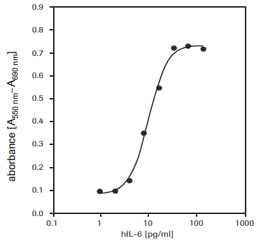
7TD1細胞の増殖

図3.組換えヒトインターロイキン-6(hIL-6)に応答した7TD1細胞(マウス-マウスハイブリドーマ)の増殖をMTTアッセイで測定

同様のアッセイ

XTTアッセイとWST-1アッセイも細胞生存率と細胞増殖を測定するために使用できます。

製品

Loading

参考文献

1.
Mosmann T. 1983. Rapid colorimetric assay for cellular growth and survival: Application to proliferation and cytotoxicity assays. Journal of Immunological Methods. 65(1-2):55-63. https://doi.org/10.1016/0022-1759(83)90303-4
2.
Tada H, Shiho O, Kuroshima K, Koyama M, Tsukamoto K. 1986. An improved colorimetric assay for interleukin 2. Journal of Immunological Methods. 93(2):157-165. https://doi.org/10.1016/0022-1759(86)90183-3
3.
Denizot F, Lang R. 1986. Rapid colorimetric assay for cell growth and survival. Journal of Immunological Methods. 89(2):271-277. https://doi.org/10.1016/0022-1759(86)90368-6
4.
Gerlier D, Thomasset N. 1986. Use of MTT colorimetric assay to measure cell activation. Journal of Immunological Methods. 94(1-2):57-63. https://doi.org/10.1016/0022-1759(86)90215-2
5.
Hansen MB, Nielsen SE, Berg K. 1989. Re-examination and further development of a precise and rapid dye method for measuring cell growth/cell kill. Journal of Immunological Methods. 119(2):203-210. https://doi.org/10.1016/0022-1759(89)90397-9
6.
Vistica VT, Skehan P, Scudiero D, Monks A, Pittman A, Boyd MR. 1991. Tetrazolium-based assays for cellular viability: a critical examination of selected parameters affecting formazan production. Cancer Res. 51(10):2515-20.
7.
Maehara Y, Anai H, Tamada R, Sugimachi K. 1987. The ATP assay is more sensitive than the succinate dehydrogenase inhibition test for predicting cell viability. European Journal of Cancer and Clinical Oncology. 23(3):273-276. https://doi.org/10.1016/0277-5379(87)90070-8
8.
Heeg K, Reimann J, Kabelitz D, Hardt C, Wagner H. 1985. A rapid colorimetric assay for the determination of IL-2-producing helper T cell frequencies. Journal of Immunological Methods. 77(2):237-246. https://doi.org/10.1016/0022-1759(85)90036-5
9.
Hooton, Gibbs J, Paetkan U C. 1985. Interaction of interleukin 2 with cells: quantitative analysis of effects.. J Immunol. 1352464–2473.
10.
Mosmann T, Fong T. 1989. Specific assays for cytokine production by T cells. Journal of Immunological Methods. 116(2):151-158. https://doi.org/10.1016/0022-1759(89)90198-1
11.
Ohno M, Abe T. 1991. Rapid colorimetric assay for the quantification of leukemia inhibitory factor (LIF) and interleukin-6 (IL-6). Journal of Immunological Methods. 145(1-2):199-203. https://doi.org/10.1016/0022-1759(91)90327-c
12.
Berg K, Hansen MB, Nielsen SE. 1988. J. Interferon Res.. 8((suppl. 1)):S. 67.
13.
Green LM, Reade JL, Ware CF. 1984. Rapid colormetric assay for cell viability: Application to the quantitation of cytotoxic and growth inhibitory lymphokines. Journal of Immunological Methods. 70(2):257-268. https://doi.org/10.1016/0022-1759(84)90190-x
14.
Espevik T, Nissen-Meyer J. 1986. A highly sensitive cell line, WEHI 164 clone 13, for measuring cytotoxic factor/tumor necrosis factor from human monocytes. Journal of Immunological Methods. 95(1):99-105. https://doi.org/10.1016/0022-1759(86)90322-4
15.
Ferrari M, Fornasiero MC, Isetta AM. 1990. MTT colorimetric assay for testing macrophage cytotoxic activity in vitro. Journal of Immunological Methods. 131(2):165-172. https://doi.org/10.1016/0022-1759(90)90187-z
16.
van de Loosdrecht AA, Nennie E, Ossenkoppele GJ, Beelen RH, Langenhuijsen MM. 1991. Cell mediated cytotoxicity against U 937 cells by human monocytes and macrophages in a modified colorimetric MTT assay. Journal of Immunological Methods. 141(1):15-22. https://doi.org/10.1016/0022-1759(91)90205-t
17.
Cole S. 1986. Rapid chemosensitivity testing of human lung tumor cells using the MTT assay. Cancer Chemother. Pharmacol.. 17(3): https://doi.org/10.1007/bf00256695
18.
Carmichael J, DeGraff WG, Gazdar AF, Minna JD, Mitchell JB. 1987. Evaluation of a tetrazolium-based semiautomated colorimetric assay: assessment of chemosensitivity testing. Cancer Res. 47(4):936-42.
19.
Twentyman P, Luscombe M. 1987. A study of some variables in a tetrazolium dye (MTT) based assay for cell growth and chemosensitivity. Br J Cancer. 56(3):279-285. https://doi.org/10.1038/bjc.1987.190
20.
Park JG, Park BS, Steinberg SM, Carmichael J, Collins JM, Minna JD, Gazdar AF. 1987. Chemosensitivity testing of human colorectal carcinoma cell lines using a tetrazolium-based colorimetric assay. Cancer Res. 475875-9.

21.
1988. FFPS News. Oryx. 22(4):246-250. https://doi.org/10.1017/s0030605300022481
22.
Pieters R, Huismans D, Leyva A, Veerman A. 1988. Adaptation of the rapid automated tetrazolium dye based (MTT) assay for chemosensitivity testing in childhood leukemia. Cancer Letters. 41(3):323-332. https://doi.org/10.1016/0304-3835(88)90294-7
23.
McHaLe A, McHaLe L. 1988. Use of a tetrazolium based colorimetric assay in assessing photoradiation therapy in vitro. Cancer Letters. 41(3):315-321. https://doi.org/10.1016/0304-3835(88)90293-5
24.
Edmondson JM, Armstrong LS, Martinez AO. 1988. A rapid and simple MTT-based spectrophotometric assay for determining drug sensitivity in monolayer cultures. Journal of Tissue Culture Methods. 11(1):15-17. https://doi.org/10.1007/bf01404408
25.
Faircloth GT, Stewart D, Clement JJ. 1988. A simple screening procedure for the quantitative measurement of cytotoxicity to resting primary lymphocyte cultures. Journal of Tissue Culture Methods. 11(4):201-205. https://doi.org/10.1007/bf01407314
26.
Campling BG, Pym J, Galbraith PR, Cole SP. 1988. Use of the mtt assay for rapid determination of chemosensitivity of human leukemic blast cells. Leukemia Research. 12(10):823-831. https://doi.org/10.1016/0145-2126(88)90036-7
27.
Pieters R, Huismans D, Leyva A, Veerman A. 1989. Comparison of the rapid automated MTT-assay with a dye exclusion assay for chemosensitivity testing in childhood leukaemia. Br J Cancer. 59(2):217-220. https://doi.org/10.1038/bjc.1989.44
28.
Twentyman PR, Fox NE, Rees JKH. 1989. Chemosensitivity testing of fresh leukaemia cells using the MTT colorimetric assay. Br J Haematol. 71(1):19-24. https://doi.org/10.1111/j.1365-2141.1989.tb06268.x
29.
Plumb JA, Milroy R, Kaye SB. 1989. Effects of the pH dependence of 3-(4,5-dimethylthiazol-2-yl)-2,5-diphenyl-tetrazolium bromide-formazan absorption on chemosensitivity determined by a novel tetrazolium-based assay. Cancer Res. 494435–4440.
30.
Alley MC, Scudiero DA, Monks A, Hursey ML, Czerwinski MJ, Fine DL, Abbott BJ, Mayo JG, Shoemaker RH, Boyd MR. 1988. Feasibility of drug screening with panels of human tumor cell lines using a microculture tetrazolium assay. Cancer Res. 48589–601.
31.
Heo DS, Park JG, Hata K, Day R, Herberman RB, Whiteside TL. 1990. Evaluation of tetrazolium-based semiautomatic colorimetric assay for measurement of human antitumor cytotoxicity. Cancer Res. 503681–3690.
32.
Ruben RL, Neubauer RH. 1987. Semiautomated colorimetric assay for in vitro screening of anticancer compounds. Cancer Treat Rep. 711141–1149..
33.
Rubinstein LV, Shoemaker RH, Paull KD, Simon RM, Tosini S, Skehan P, Scudiero DA, Monks A, Boyd MR. 1990. Comparison of In Vitro Anticancer-Drug-Screening Data Generated With a Tetrazolium Assay Versus a Protein Assay Against a Diverse Panel of Human Tumor Cell Lines. JNCI Journal of the National Cancer Institute. 82(13):1113-1117. https://doi.org/10.1093/jnci/82.13.1113
34.
1991. Announcement. Utilitas. 3(2):330-330. https://doi.org/10.1017/s0953820800001254
35.
Slater T, Sawyer B, Sträuli U. 1963. Studies on succinate-tetrazolium reductase systems. Biochimica et Biophysica Acta. 77383-393. https://doi.org/10.1016/0006-3002(63)90513-4
36.
Berridge M, Tan A. 1993. Characterization of the Cellular Reduction of 3-(4,5-dimethylthiazol-2-yl)-2,5-diphenyltetrazolium bromide (MTT): Subcellular Localization, Substrate Dependence, and Involvement of Mitochondrial Electron Transport in MTT Reduction. Archives of Biochemistry and Biophysics. 303(2):474-482. https://doi.org/10.1006/abbi.1993.1311