
S7100
ApopTagペルオキシダーゼIn Situアポトーシス検出キット
The ApopTag Peroxidase In Situ Apoptosis Detection Kit detects apoptotic cells in situ by labeling & detecting DNA strand breaks by the TUNEL method.
別名:
ApopTag キット, In Situ アポトーシス検出キット
manufacturer/tradename
ApopTag, Chemicon®
detection method
colorimetric
shipped in
dry ice
Quality Level
General description
Of all the aspects of apoptosis, the defining characteristic is a complete change in cellular morphology. As observed by electron microscopy, the cell undergoes shrinkage, chromatin margination, membrane blebbing, nuclear condensation and then segmentation, and division into apoptotic bodies which may be phagocytosed (11, 19, 24). The characteristic apoptotic bodies are short-lived and minute, and can resemble other cellular constituents when viewed by brightfield microscopy. DNA fragmentation in apoptotic cells is followed by cell death and removal from the tissue, usually within several hours (7). A rate of tissue regression as rapid as 25% per day can result from apparent apoptosis in only 2-3% of the cells at any one time (6). Thus, the quantitative measurement of an apoptotic index by morphology alone can be difficult.
DNA fragmentation is usually associated with ultrastructural changes in cellular morphology in apoptosis (26, 38). In a number of well-researched model systems, large fragments of 300 kb and 50 kb are first produced by endonucleolytic degradation of higher-order chromatin structural organization. These large DNA fragments are visible on pulsed-field electrophoresis gels (5, 43, 44). In most models, the activation of Ca2+- and Mg2+-dependent endonuclease activity further shortens the fragments by cleaving the DNA at linker sites between nucleosomes (3). The ultimate DNA fragments are multimers of about 180 bp nucleosomal units. These multimers appear as the familiar "DNA ladder" seen on standard agarose electrophoresis gels of DNA extracted from many kinds of apoptotic cells (e.g. 3, 7,13, 35, 44).
Another method for examining apoptosis via DNA fragmentation is by the TUNEL assay, (13) which is the basis of ApopTag technology. The DNA strand breaks are detected by enzymatically labeling the free 3′-OH termini with modified nucleotides. These new DNA ends that are generated upon DNA fragmentation are typically localized in morphologically identifiable nuclei and apoptotic bodies. In contrast, normal or proliferative nuclei, which have relatively insignificant numbers of DNA 3′-OH ends, usually do not stain with the kit. ApopTag Kits detect single-stranded (25) and double-stranded breaks associated with apoptosis. Drug-induced DNA damage is not identified by the TUNEL assay unless it is coupled to the apoptotic response (8). In addition, this technique can detect early-stage apoptosis in systems where chromatin condensation has begun and strand breaks are fewer, even before the nucleus undergoes major morphological changes (4, 8).
Apoptosis is distinct from accidental cell death (necrosis). Numerous morphological and biochemical differences that distinguish apoptotic from necrotic cell death are summarized in the following table (adapted with permission from reference 39).
Preparation Note
使用上の注意
1. 以下のキット構成要素には、バッファーとしてカコジル酸カリウム(ジメチルアルシン酸)が含まれています:平衡バッファー(#90416)、反応バッファー(#90417)、TdT酵素(#90418)。これらの構成要素は飲み込むと有害です。皮膚と眼への接触を避け(手袋、眼鏡を着用)、接触した部位はすぐに洗ってください。
2.抗体コンジュゲート(90420)およびブロッキング溶液(10および13)には、保存剤として0.08%アジ化ナトリウムが含まれています。
3.TdT酵素(#90418)にはグリセロールが含まれており、-20°Cでは凍りません。有効期間を最長にするため、分注する前にこの試薬が室温まで温まらないようにしてください。
Other Notes
反応バッファー 2.0 mL -15°C~-25°C
TdT酵素 0.64 mL -15°C~-25°C
停止/洗浄バッファー 20 mL -15°C~-25°C
抗ジゴキシゲニン-ペルオキシダーゼ* 3.0 mL 2°C~8°C
プラスチックカバースリップ 100 ea.室温。
注記:DAB(ペルオキシダーゼ基質)は別途購入する必要があります。このキットには付属していません。
1キットあたりのサンプル数:指示に従って使用する場合は、約5 mm2の組織サンプル40点分の染色に十分な材料が含まれています。キットをスライド標本に使用する場合は、他の試薬より先に反応バッファーが完全に消費されます。
Legal Information
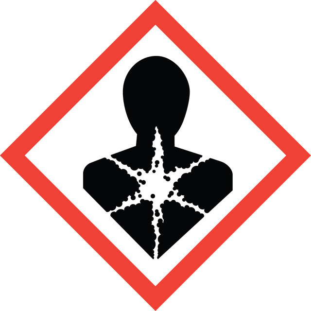

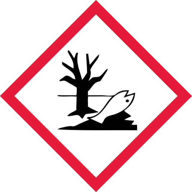
signalword
Danger
hcodes
Hazard Classifications
Aquatic Chronic 2 - Carc. 1B - Skin Sens. 1 - STOT RE 2 Inhalation
target_organs
Respiratory Tract
保管分類
6.1C - Combustible acute toxic Cat.3 / toxic compounds or compounds which causing chronic effects
wgk
WGK 3
適用法令
試験研究用途を考慮した関連法令を主に挙げております。化学物質以外については、一部の情報のみ提供しています。 製品を安全かつ合法的に使用することは、使用者の義務です。最新情報により修正される場合があります。WEBの反映には時間を要することがあるため、適宜SDSをご参照ください。
キットコンポーネントの情報を参照してください
pdsc
キットコンポーネントの情報を参照してください
prtr
キットコンポーネントの情報を参照してください
fsl
キットコンポーネントの情報を参照してください
ishl_indicated
キットコンポーネントの情報を参照してください
ishl_notified
キットコンポーネントの情報を参照してください
cart
キットコンポーネントの情報を参照してください
jan
試験成績書(COA)
製品のロット番号・バッチ番号を入力して、試験成績書(COA) を検索できます。ロット番号・バッチ番号は、製品ラベルに「Lot」または「Batch」に続いて記載されています。
資料
Cellular apoptosis assays to detect programmed cell death using Annexin V, Caspase and TUNEL DNA fragmentation assays.
関連コンテンツ
"Recognizing both the tremendous opportunities and the challenges facing cancer research, we are dedicated to developing and refining tools and technologies for the study of cancer. With our comprehensive portfolio, including the Upstate®, Chemicon®, and Calbiochem® brands of reagents and antibodies, researchers can count on dependable, high quality solutions for analyzing all the hallmarks of cancer."
グローバルトレードアイテム番号
| カタログ番号 | GTIN |
|---|---|
| S7100 | 04053252579356 |
ライフサイエンス、有機合成、材料科学、クロマトグラフィー、分析など、あらゆる分野の研究に経験のあるメンバーがおります。.
製品に関するお問い合わせはこちら(テクニカルサービス)