Wenn Sie das Fenster schließen, wird Ihre Konfiguration nicht gespeichert, es sei denn, Sie haben Ihren Artikel in die Bestellung aufgenommen oder zu Ihren Favoriten hinzugefügt.
Klicken Sie auf OK, um das MILLIPLEX® MAP-Tool zu schließen oder auf Abbrechen, um zu Ihrer Auswahl zurückzukehren.
Wählen Sie konfigurierbare Panels & Premixed-Kits - ODER - Kits für die zelluläre Signaltransduktion & MAPmates™
Konfigurieren Sie Ihre MILLIPLEX® MAP-Kits und lassen sich den Preis anzeigen.
Konfigurierbare Panels & Premixed-Kits
Unser breites Angebot enthält Multiplex-Panels, für die Sie die Analyten auswählen können, die am besten für Ihre Anwendung geeignet sind. Unter einem separaten Register können Sie das Premixed-Cytokin-Format oder ein Singleplex-Kit wählen.
Kits für die zelluläre Signaltransduktion & MAPmates™
Wählen Sie gebrauchsfertige Kits zur Erforschung gesamter Signalwege oder Prozesse. Oder konfigurieren Sie Ihre eigenen Kits mit Singleplex MAPmates™.
Die folgenden MAPmates™ sollten nicht zusammen analysiert werden: -MAPmates™, die einen unterschiedlichen Assaypuffer erfordern. -Phosphospezifische und MAPmate™ Gesamtkombinationen wie Gesamt-GSK3β und Gesamt-GSK3β (Ser 9). -PanTyr und locusspezifische MAPmates™, z.B. Phospho-EGF-Rezeptor und Phospho-STAT1 (Tyr701). -Mehr als 1 Phospho-MAPmate™ für ein einziges Target (Akt, STAT3). -GAPDH und β-Tubulin können nicht mit Kits oder MAPmates™, die panTyr enthalten, analysiert werden.
.
Bestellnummer
Bestellinformationen
St./Pkg.
Liste
Dieser Artikel wurde zu Ihren Favoriten hinzugefügt.
Wählen Sie bitte Spezies, Panelart, Kit oder Probenart
Um Ihr MILLIPLEX® MAP-Kit zu konfigurieren, wählen Sie zunächst eine Spezies, eine Panelart und/oder ein Kit.
Custom Premix Selecting "Custom Premix" option means that all of the beads you have chosen will be premixed in manufacturing before the kit is sent to you.
Catalogue Number
Ordering Description
Qty/Pack
List
Dieser Artikel wurde zu Ihren Favoriten hinzugefügt.
Spezies
Panelart
Gewähltes Kit
Menge
Bestellnummer
Bestellinformationen
St./Pkg.
Listenpreis
96-Well Plate
Menge
Bestellnummer
Bestellinformationen
St./Pkg.
Listenpreis
Weitere Reagenzien hinzufügen (MAPmates erfordern die Verwendung eines Puffer- und Detektionskits)
Menge
Bestellnummer
Bestellinformationen
St./Pkg.
Listenpreis
48-602MAG
Buffer Detection Kit for Magnetic Beads
1 Kit
Platzsparende Option Kunden, die mehrere Kits kaufen, können ihre Multiplex-Assaykomponenten in Kunststoffbeuteln anstelle von Packungen erhalten, um eine kompaktere Lagerung zu ermöglichen.
Dieser Artikel wurde zu Ihren Favoriten hinzugefügt.
Das Produkt wurde in Ihre Bestellung aufgenommen
Sie können nun ein weiteres Kit konfigurieren, ein Premixed-Kit wählen, zur Kasse gehen oder das Bestell-Tool schließen.
The goal of drug discovery is to find well-qualified candidate drugs expected to provide therapeutic benefit. Use of relevant and information-rich assays can rapidly eliminate candidates that either do not affect the appropriate response or cause some unforeseen lethality, and can save drug companies time and money that is often wasted pursuing clinical trials with flawed drug candidates. Many of the Amnis® imaging flow cytometry's applications are ideally suited for characterizing drug candidates, including cell signaling, cell-cell interactions, chemokine-induced shape change, cell death, co-localization, and phagocytosis, to name a few. Furthermore, these applications work with primary cells such as whole blood leukocytes and can often be multiplexed.
Co-Localization of an Antibody-Drug Conjugate to Endosomes or Lysosomes
In this experiment we use the capabilities of the Imagestream®x system to measure the transit of a fluorescently labeled antibody-drug conjugate (ADC) through the cellular endocytic pathway. The high spatial resolution afforded by the Imagestream®x system allowed accurate measurement of ADC co-localization to endosomes and lysosomes. See the application note for more details.
Barcoding
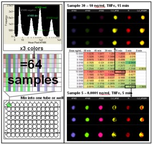
Screening drug candidates requires the rapid analysis of large numbers of samples. Multiplexing samples on the Imagestream®x by mixing dozens of samples from a time course or dose response titration into one tube dramatically increases the sample throughput of the system. This is achieved through fluorescent cell barcoding with subsequent identification of each sample based on a pattern of fluorescent dye and staining intensity unique to each treatment/time point. Automated location of populations based on fluorescence intensity and color allows deconvolution of the barcoded samples after acquisition.
Here is an example of measuring the movement of NF-κB from the cytoplasm to the nucleus in response to varying doses and times of exposure to TNF-α. By labeling 64 separate samples with different combinations of 3 dyes (AlexaFluor 405, 488, and 660) at 4 intensity levels (43 = 64) it was possible to run one tube of the combined samples on the Imagestream®x and acquire a single file. After acquisition of over 600,000 images in 10-15 minutes, the color and intensity combinations of the samples were deconvoluted for further quantitative image analysis. Mean Similarity scores are reported in the spreadsheet and a heatmap has been applied. See the cell signaling application for more information about the translocation assay. The combination of high speed image acquisition and automated quantitative image analysis provided by the Imagestream®x system offers a major advancement in the field of drug discovery.