Wenn Sie das Fenster schließen, wird Ihre Konfiguration nicht gespeichert, es sei denn, Sie haben Ihren Artikel in die Bestellung aufgenommen oder zu Ihren Favoriten hinzugefügt.
Klicken Sie auf OK, um das MILLIPLEX® MAP-Tool zu schließen oder auf Abbrechen, um zu Ihrer Auswahl zurückzukehren.
Wählen Sie konfigurierbare Panels & Premixed-Kits - ODER - Kits für die zelluläre Signaltransduktion & MAPmates™
Konfigurieren Sie Ihre MILLIPLEX® MAP-Kits und lassen sich den Preis anzeigen.
Konfigurierbare Panels & Premixed-Kits
Unser breites Angebot enthält Multiplex-Panels, für die Sie die Analyten auswählen können, die am besten für Ihre Anwendung geeignet sind. Unter einem separaten Register können Sie das Premixed-Cytokin-Format oder ein Singleplex-Kit wählen.
Kits für die zelluläre Signaltransduktion & MAPmates™
Wählen Sie gebrauchsfertige Kits zur Erforschung gesamter Signalwege oder Prozesse. Oder konfigurieren Sie Ihre eigenen Kits mit Singleplex MAPmates™.
Die folgenden MAPmates™ sollten nicht zusammen analysiert werden: -MAPmates™, die einen unterschiedlichen Assaypuffer erfordern. -Phosphospezifische und MAPmate™ Gesamtkombinationen wie Gesamt-GSK3β und Gesamt-GSK3β (Ser 9). -PanTyr und locusspezifische MAPmates™, z.B. Phospho-EGF-Rezeptor und Phospho-STAT1 (Tyr701). -Mehr als 1 Phospho-MAPmate™ für ein einziges Target (Akt, STAT3). -GAPDH und β-Tubulin können nicht mit Kits oder MAPmates™, die panTyr enthalten, analysiert werden.
.
Bestellnummer
Bestellinformationen
St./Pkg.
Liste
Dieser Artikel wurde zu Ihren Favoriten hinzugefügt.
Wählen Sie bitte Spezies, Panelart, Kit oder Probenart
Um Ihr MILLIPLEX® MAP-Kit zu konfigurieren, wählen Sie zunächst eine Spezies, eine Panelart und/oder ein Kit.
Custom Premix Selecting "Custom Premix" option means that all of the beads you have chosen will be premixed in manufacturing before the kit is sent to you.
Catalogue Number
Ordering Description
Qty/Pack
List
Dieser Artikel wurde zu Ihren Favoriten hinzugefügt.
Spezies
Panelart
Gewähltes Kit
Menge
Bestellnummer
Bestellinformationen
St./Pkg.
Listenpreis
96-Well Plate
Menge
Bestellnummer
Bestellinformationen
St./Pkg.
Listenpreis
Weitere Reagenzien hinzufügen (MAPmates erfordern die Verwendung eines Puffer- und Detektionskits)
Menge
Bestellnummer
Bestellinformationen
St./Pkg.
Listenpreis
48-602MAG
Buffer Detection Kit for Magnetic Beads
1 Kit
Platzsparende Option Kunden, die mehrere Kits kaufen, können ihre Multiplex-Assaykomponenten in Kunststoffbeuteln anstelle von Packungen erhalten, um eine kompaktere Lagerung zu ermöglichen.
Dieser Artikel wurde zu Ihren Favoriten hinzugefügt.
Das Produkt wurde in Ihre Bestellung aufgenommen
Sie können nun ein weiteres Kit konfigurieren, ein Premixed-Kit wählen, zur Kasse gehen oder das Bestell-Tool schließen.
Anti-CD6, clone UMCD6, Cat. No. MABF2105 is a mouse monoclonal antibody that detects CD6 and has been tested for use in Dot Blot, Flow Cytometry, Immunoprecipitation, Function Analysis, Inhibition assay.
More>>Anti-CD6, clone UMCD6, Cat. No. MABF2105 is a mouse monoclonal antibody that detects CD6 and has been tested for use in Dot Blot, Flow Cytometry, Immunoprecipitation, Function Analysis, Inhibition assay. Less<<
Anti-CD6 Antibody, clone UMCD6: SDB (Sicherheitsdatenblätter), Analysenzertifikate und Qualitätszertifikate, Dossiers, Broschüren und andere verfügbare Dokumente.
T-cell differentiation antigen CD6 (UniProt: P30203; also known as T12, TP120, CD6) is encoded by the CD6 gene (Gene ID: 923) in human. CD6 is a single-pass type I membrane protein that serves as a cell adhesion molecule that mediates cell-cell contacts and regulates T-cell responses via its interaction with ALCAM/CD166. It contributes to signaling cascades triggered by activation of the TCR/CD3 complex and functions as a costimulatory molecule to promote T-cell activation and proliferation. It is also involved in the formation and maturation of the immunological synapse. CD6 has been detected on peripheral blood T-cells and on natural killer (NK) cells and is also detected in spleen, thymus, appendix, lymph nodes. CD6 is known to bind both lipopolysaccharide (LPS) from Gram-negative bacteria and lipoteichoic acid from Gram-positive bacteria and LPS binding leads to the activation of signaling cascades and down-stream MAP kinases. It is synthesized with a signal peptide (aa 1-17), which is subsequently cleaved off in the mature form. It has an extracellular domain (aa 18-402), a transmembrane domain (aa 403-423), and a cytoplasmic domain (aa 424-668). Seven isoforms of CD6 have been reported that are produced by alternative splicing.
References
Product Information
Format
Purified
Presentation
Purified mouse monoclonal antibody IgG1 in PBS without preservatives.
Applications
Application
Anti-CD6, clone UMCD6, Cat. No. MABF2105 is a mouse monoclonal antibody that detects CD6 and has been tested for use in Dot Blot, Flow Cytometry, Immunoprecipitation, Function Analysis, Inhibition assay.
Key Applications
Dot Blot
Flow Cytometry
Immunoprecipitation
Function Assay
Inhibition
Application Notes
Inhibition Analysis: A representative lot inhibited antigen-specific responses to tetanus toxoid, but not to the superantigen SEA. (Singer, N.G., et. al. (1996). Immunology. 88(4):537-43).
Induces Function: A representative lot showed strong synergistic effect with phorbol ester in inducing T cell activation and enhanced the autologous mixed lymphocyte reaction. (Bott, C.M., et. al. (1993). Int Immunol. 5(7):783-92).
Dot Blot Analysis: A representative lot detected CD6 in Dot Blot applications (Bott, C.M., et. al. (1993). Int Immunol. 5(7):783-92).
Immunoprecipitation Analysis: A representative lot immunoprecipitated CD6 in Immunoprecipitation applications (Bott, C.M., et. al. (1993). Int Immunol. 5(7):783-92).
Flow Cytometry Analysis: A representative lot detected CD6 in Immunoprecipitation applications (Singer, N.G., et. al. (1996). Immunology. 88(4):537-43; Bott, C.M., et. al. (1993). Int Immunol. 5(7):783-92; Bott, C.M., et. al. (1994). J Immunol. 153(1):1-9; Li, Y., et. al. (2017). Proc Natl Acad Sci USA. 114(10):2687-2692).
Biological Information
Immunogen
ST-1 T cells isolated from human knee synovium.
Clone
UMCD6
Concentration
Please refer to lot specific datasheet.
Host
Mouse
Specificity
Clone UMCD6 specifically detects CD6 in human cells.
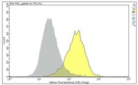
Evaluated by Flow Cytometry in PMA stimulated Jurkat cells.
Flow Cytometry Analysis: A 1:1000 dilution of this antibody detected CD6 in one million Jurkat cells stimulated with PMA.
Usage Statement
Unless otherwise stated in our catalog or other company documentation accompanying the product(s), our products are intended for research use only and are not to be used for any other purpose, which includes but is not limited to, unauthorized commercial uses, in vitro diagnostic uses, ex vivo or in vivo therapeutic uses or any type of consumption or application to humans or animals.
Storage and Shipping Information
Storage Conditions
Stable for 1 year at -20°C from date of receipt. Handling Recommendations: Upon receipt and prior to removing the cap, centrifuge the vial and gently mix the solution. Aliquot into microcentrifuge tubes and store at -20°C. Avoid repeated freeze/thaw cycles, which may damage IgG and affect product performance.