Wenn Sie das Fenster schließen, wird Ihre Konfiguration nicht gespeichert, es sei denn, Sie haben Ihren Artikel in die Bestellung aufgenommen oder zu Ihren Favoriten hinzugefügt.
Klicken Sie auf OK, um das MILLIPLEX® MAP-Tool zu schließen oder auf Abbrechen, um zu Ihrer Auswahl zurückzukehren.
Wählen Sie konfigurierbare Panels & Premixed-Kits - ODER - Kits für die zelluläre Signaltransduktion & MAPmates™
Konfigurieren Sie Ihre MILLIPLEX® MAP-Kits und lassen sich den Preis anzeigen.
Konfigurierbare Panels & Premixed-Kits
Unser breites Angebot enthält Multiplex-Panels, für die Sie die Analyten auswählen können, die am besten für Ihre Anwendung geeignet sind. Unter einem separaten Register können Sie das Premixed-Cytokin-Format oder ein Singleplex-Kit wählen.
Kits für die zelluläre Signaltransduktion & MAPmates™
Wählen Sie gebrauchsfertige Kits zur Erforschung gesamter Signalwege oder Prozesse. Oder konfigurieren Sie Ihre eigenen Kits mit Singleplex MAPmates™.
Die folgenden MAPmates™ sollten nicht zusammen analysiert werden: -MAPmates™, die einen unterschiedlichen Assaypuffer erfordern. -Phosphospezifische und MAPmate™ Gesamtkombinationen wie Gesamt-GSK3β und Gesamt-GSK3β (Ser 9). -PanTyr und locusspezifische MAPmates™, z.B. Phospho-EGF-Rezeptor und Phospho-STAT1 (Tyr701). -Mehr als 1 Phospho-MAPmate™ für ein einziges Target (Akt, STAT3). -GAPDH und β-Tubulin können nicht mit Kits oder MAPmates™, die panTyr enthalten, analysiert werden.
.
Bestellnummer
Bestellinformationen
St./Pkg.
Liste
Dieser Artikel wurde zu Ihren Favoriten hinzugefügt.
Wählen Sie bitte Spezies, Panelart, Kit oder Probenart
Um Ihr MILLIPLEX® MAP-Kit zu konfigurieren, wählen Sie zunächst eine Spezies, eine Panelart und/oder ein Kit.
Custom Premix Selecting "Custom Premix" option means that all of the beads you have chosen will be premixed in manufacturing before the kit is sent to you.
Catalogue Number
Ordering Description
Qty/Pack
List
Dieser Artikel wurde zu Ihren Favoriten hinzugefügt.
Spezies
Panelart
Gewähltes Kit
Menge
Bestellnummer
Bestellinformationen
St./Pkg.
Listenpreis
96-Well Plate
Menge
Bestellnummer
Bestellinformationen
St./Pkg.
Listenpreis
Weitere Reagenzien hinzufügen (MAPmates erfordern die Verwendung eines Puffer- und Detektionskits)
Menge
Bestellnummer
Bestellinformationen
St./Pkg.
Listenpreis
48-602MAG
Buffer Detection Kit for Magnetic Beads
1 Kit
Platzsparende Option Kunden, die mehrere Kits kaufen, können ihre Multiplex-Assaykomponenten in Kunststoffbeuteln anstelle von Packungen erhalten, um eine kompaktere Lagerung zu ermöglichen.
Dieser Artikel wurde zu Ihren Favoriten hinzugefügt.
Das Produkt wurde in Ihre Bestellung aufgenommen
Sie können nun ein weiteres Kit konfigurieren, ein Premixed-Kit wählen, zur Kasse gehen oder das Bestell-Tool schließen.
Anti-CP110, clone 140-195-5 , Cat. No. MABT1354, is a mouse monoclonal antibody that detects Centriolar coiled-coil protein of 110 kDa and has been tested for use in immunocytochemistry, Immunofluorescence, Immunoprecipitation, and Western Blotting.
More>>Anti-CP110, clone 140-195-5 , Cat. No. MABT1354, is a mouse monoclonal antibody that detects Centriolar coiled-coil protein of 110 kDa and has been tested for use in immunocytochemistry, Immunofluorescence, Immunoprecipitation, and Western Blotting. Less<<
Anti-CP110 Antibody, clone 140-195-5: SDB (Sicherheitsdatenblätter), Analysenzertifikate und Qualitätszertifikate, Dossiers, Broschüren und andere verfügbare Dokumente.
Centriolar coiled-coil protein of 110 kDa (UniProt: O43303; also known as Centrosomal protein of 110 kDa, CP110, Cep110) is encoded by the CCP110 (also known as CEP110, CP110, KIAA0419) gene (Gene ID: 9738) in human. CP110 is a cytoplasmic protein that is essential for centrosome duplication at different stages of procentriole formation. It acts as a key negative regulator of ciliogenesis in collaboration with CEP97 by capping the mother centriole thereby preventing cilia formation. It is also involved in the assembly of the mother centriole subdistal appendages thereby affecting the fusion of recycling endosomes to basal bodies during cilia formation. Two isoforms of CP110 have been reported that are produced by alternative splicing. CP110 is highly expressed in testis and intermediate level of expression is observed in spleen, thymus, prostate, small intestine, colon and peripheral blood leukocytes. CP110 levels re up-regulated during the transition from G1 to S phase of the cell cycle and highest levels are reported in S phase, following which the levels decline significantly. During the G2 phase it is ubiquitinated that leads to its proteasomal degradation, which prevents centrosome reduplication. And, de-ubiquitinated by USP33 in S and G2/M phase stabilizes CCP110 during the period when centrioles duplicate and elongate.
References
Product Information
Format
Purified
Presentation
Purified mouse monoclonal antibody IgG1 in buffer containing 0.1 M Tris-Glycine (pH 7.4), 150 mM NaCl with 0.05% sodium azide.
Anti-CP110, clone 140-195-5 , Cat. No. MABT1354, is a mouse monoclonal antibody that detects Centriolar coiled-coil protein of 110 kDa and has been tested for use in immunocytochemistry, Immunofluorescence, Immunoprecipitation, and Western Blotting.
Key Applications
Immunocytochemistry
Immunofluorescence
Immunoprecipitation
Western Blotting
Application Notes
Immunoprecipitation Analysis: A representative lot detected CP110 in HEK293T cell lysate (Courtesy of Prof. Erich A. Nigg, Biozentrum, University of Basel, Switzerland).
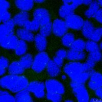
Immunocytochemistry Analysis: A 1:50 dilution from a representative lot detected CP110 in U2OS cell line.
Western Blotting Analysis: A representative lot detected CP110 in Western Blotting applications (Bauer, M., et. al. (2016). EMBO J. 35(19):2152-2166).
Immunofluorescence Analysis: A representative lot detected CP110 in U20S cells (Courtesy of Prof. Erich A. Nigg, Biozentrum, University of Basel, Switzerland).
Western Blotting Analysis: A representative lot detected CP110 in 293T cell lysate (Courtesy of Prof. Erich A. Nigg, Biozentrum, University of Basel, Switzerland).
Immunofluorescence Analysis: A representative lot detected CP110 in Western Blotting applications (Bauer, M., et. al. (2016). EMBO J. 35(19):2152-2166; Arquint, C., et. al. (2014). Curr Biol. 24(4):351-60).
Biological Information
Immunogen
His-tagged recombinant fragment corresponding to the first 149 amino acids from the N-terminal region of human Centriolar coiled-coil protein of 110 kDa.
Epitope
N-terminus
Clone
140-195-5
Concentration
Please refer to lot specific datasheet.
Host
Mouse
Specificity
Clone 140-195-5 detects Centriolar coiled-coil protein of 110 kDa (CP110) in human cells., It targets an epitope within the first 149 amino acids from the N-termina region.
~150 kDa observed; 113.42 kDa calculated. Uncharacterized bands may be observed in some lysate(s).
Physicochemical Information
Dimensions
Materials Information
Toxicological Information
Safety Information according to GHS
Safety Information
Product Usage Statements
Quality Assurance
Evaluated by Western Blotting in HEK293 cell lysate.
Western Blotting Analysis: A 1:250 dilution of this antibody detected CP110 in HEK293 cell lysate.
Usage Statement
Unless otherwise stated in our catalog or other company documentation accompanying the product(s), our products are intended for research use only and are not to be used for any other purpose, which includes but is not limited to, unauthorized commercial uses, in vitro diagnostic uses, ex vivo or in vivo therapeutic uses or any type of consumption or application to humans or animals.






 Antibody[198945-ALL].jpg)
