Wenn Sie das Fenster schließen, wird Ihre Konfiguration nicht gespeichert, es sei denn, Sie haben Ihren Artikel in die Bestellung aufgenommen oder zu Ihren Favoriten hinzugefügt.
Klicken Sie auf OK, um das MILLIPLEX® MAP-Tool zu schließen oder auf Abbrechen, um zu Ihrer Auswahl zurückzukehren.
Wählen Sie konfigurierbare Panels & Premixed-Kits - ODER - Kits für die zelluläre Signaltransduktion & MAPmates™
Konfigurieren Sie Ihre MILLIPLEX® MAP-Kits und lassen sich den Preis anzeigen.
Konfigurierbare Panels & Premixed-Kits
Unser breites Angebot enthält Multiplex-Panels, für die Sie die Analyten auswählen können, die am besten für Ihre Anwendung geeignet sind. Unter einem separaten Register können Sie das Premixed-Cytokin-Format oder ein Singleplex-Kit wählen.
Kits für die zelluläre Signaltransduktion & MAPmates™
Wählen Sie gebrauchsfertige Kits zur Erforschung gesamter Signalwege oder Prozesse. Oder konfigurieren Sie Ihre eigenen Kits mit Singleplex MAPmates™.
Die folgenden MAPmates™ sollten nicht zusammen analysiert werden: -MAPmates™, die einen unterschiedlichen Assaypuffer erfordern. -Phosphospezifische und MAPmate™ Gesamtkombinationen wie Gesamt-GSK3β und Gesamt-GSK3β (Ser 9). -PanTyr und locusspezifische MAPmates™, z.B. Phospho-EGF-Rezeptor und Phospho-STAT1 (Tyr701). -Mehr als 1 Phospho-MAPmate™ für ein einziges Target (Akt, STAT3). -GAPDH und β-Tubulin können nicht mit Kits oder MAPmates™, die panTyr enthalten, analysiert werden.
.
Bestellnummer
Bestellinformationen
St./Pkg.
Liste
Dieser Artikel wurde zu Ihren Favoriten hinzugefügt.
Wählen Sie bitte Spezies, Panelart, Kit oder Probenart
Um Ihr MILLIPLEX® MAP-Kit zu konfigurieren, wählen Sie zunächst eine Spezies, eine Panelart und/oder ein Kit.
Custom Premix Selecting "Custom Premix" option means that all of the beads you have chosen will be premixed in manufacturing before the kit is sent to you.
Catalogue Number
Ordering Description
Qty/Pack
List
Dieser Artikel wurde zu Ihren Favoriten hinzugefügt.
Spezies
Panelart
Gewähltes Kit
Menge
Bestellnummer
Bestellinformationen
St./Pkg.
Listenpreis
96-Well Plate
Menge
Bestellnummer
Bestellinformationen
St./Pkg.
Listenpreis
Weitere Reagenzien hinzufügen (MAPmates erfordern die Verwendung eines Puffer- und Detektionskits)
Menge
Bestellnummer
Bestellinformationen
St./Pkg.
Listenpreis
48-602MAG
Buffer Detection Kit for Magnetic Beads
1 Kit
Platzsparende Option Kunden, die mehrere Kits kaufen, können ihre Multiplex-Assaykomponenten in Kunststoffbeuteln anstelle von Packungen erhalten, um eine kompaktere Lagerung zu ermöglichen.
Dieser Artikel wurde zu Ihren Favoriten hinzugefügt.
Das Produkt wurde in Ihre Bestellung aufgenommen
Sie können nun ein weiteres Kit konfigurieren, ein Premixed-Kit wählen, zur Kasse gehen oder das Bestell-Tool schließen.
Anti-Caspase-8, clone C15, Cat. No. MABS2498, is a mouse monoclonal antibody that detects Caspase-8 and is tested for use in Western Blotting and Immunoprecipitation.
More>>Anti-Caspase-8, clone C15, Cat. No. MABS2498, is a mouse monoclonal antibody that detects Caspase-8 and is tested for use in Western Blotting and Immunoprecipitation. Less<<
Empfohlene Produkte
Übersicht
Replacement Information
Description
Catalogue Number
MABS2498-100UG
Description
Anti-Caspase-8 Antibody, clone C15
Alternate Names
EC:3.4.22.61
CASP-8
Apoptotic cysteine protease
Apoptotic protease Mch-5
CAP4
FADD-homologous ICE/ced-3-like protease
FADD-like ICE (FLICE)
Background Information
Caspase-8 (UniProt: Q14790; also known as CASP-8, EC:3.4.22.61, Apoptotic cysteine protease, Apoptotic protease Mch-5, CAP4, FADD-homologous ICE/ced-3-like protease, FADD-like ICE (FLICE)) is encoded by the CASP8 (also known as MCH5) gene (Gene ID: 841) in human. Caspase-8 is a member of a family of cysteine-requiring aspartate proteases that participate in the intracellular signaling cascade leading to apoptosis, or programmed cell death. Caspase-8 is recruited to death-inducing signaling complex (DISC), CD95 (Fas/APO-1) and tumor necrosis factor receptor 1 (TNFR1) and is therefore the most upstream caspase in the CD95 apoptotic pathway. Caspase-8 is ubiquitously expressed in various tissues, with highest expression in peripheral blood leukocytes, spleen, thymus and liver. Its barely detectable in brain, testis and skeletal muscle. It is mainly localized in the cytoplasm and upon activation, it can translocate to the membrane-associated DISC, where it undergoes proteolytic activation. It is synthesized as an inactive proenzyme composed of an N-terminal prodomain (aa 1-216) containing two death effector domains (DEDs), followed by a large (p18) (aa 217-374) and a small (p10) (aa 385-479) catalytic subunit. Caspase-8 that binds to the death effector domain (DED) of FADD through an analogous DED domain present in the proform of caspase-8. A total of 9 isoforms are known to be produced by alternative splicing. Studies show that only two of the FLICE isoforms (caspase-8/a and caspase-8/b) were predominantly expressed in cells of different origin. Both isoforms were recruited to the CD95 death-inducing signaling complex and were activated upon CD95 stimulation with similar kinetics. (Ref.: Scaffidi, C., et al. (1997). J Biol Chem. 272(43):26953-8; Tang, D., et al. (2000). J Biol Chem. 275(13):9303-7; Teitz, T., et al. (2000). Nat Med. 6(5):529-35; Chang, D.W., et al. (2002). EMBO J. 21(14):3704-14; Lavrik, I., et al. (2003). Cell Death Differ. 10(1):144-5; Schmitz, I., et al. (2004). J Immunol. 172(4):2194-200; Lavrik, I.N., et al. (2008). J Biol Chem. 283(39):26401-8; Kallenberger, S.M., et al. (2014). Sci Signal. 7(316):ra23; de Reuver, R. et al. (2022). Nature. 607(7920):784-789).
References
Product Information
Format
Purified
Presentation
Purified mouse monoclonal antibody IgG2b in buffer containing 0.1 M Tris-Glycine (pH 7.4), 150 mM NaCl with 0.05% sodium azide.
Anti-Caspase-8, clone C15, Cat. No. MABS2498, is a mouse monoclonal antibody that detects Caspase-8 and is tested for use in Western Blotting and Immunoprecipitation.
Key Applications
Western Blotting
Immunoprecipitation
Application Notes
Tested Applications
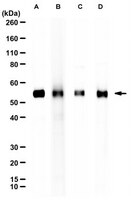
Western Blotting Analysis: A 1:1000 dilution of this antibody detected caspase-8 in Hep-G2, Jurkat, and Raji cell lysates.
Western Blotting Analysis: A representative lot detected Caspase-8 in Western Blotting applications (Scaffidi, C., et al. (1997). J Biol Chem. 272(43):26953-8; Tang, D., et al. (2000). J Biol Chem. 275(13):9303-7; Teitz, T., et al. (2000). Nat Med. 6(5):529-35; Chang, D.W., et al. (2002). EMBO J. 21(14):3704-14; Lavrik, I., et al. (2003). Cell Death Differ. 10(1):144-5; Schmitz, I., et al. (2004). J Immunol. 172(4):2194-200; Lavrik, I.N., et al. (2008). J Biol Chem. 283(39):26401-8; Kallenberger, S.M., et al. (2014). Sci Signal. 7(316):ra23; de Reuver, R. et al. (2022). Nature. 607(7920):784-789).
Immunoprecipitation Analysis: A representative lot detected Caspase-8 in Immunoprecipitation (Scaffidi, C., et al. (1997). J Biol Chem. 272(43):26953-8; Tang, D., et al. (2000). J Biol Chem. 275(13):9303-7; Teitz, T., et al. (2000). Nat Med. 6(5):529-35; Chang, D.W., et al. (2002). EMBO J. 21(14):3704-14; Lavrik, I., et al. (2003). Cell Death Differ. 10(1):144-5; Schmitz, I., et al. (2004). J Immunol. 172(4):2194-200; Lavrik, I.N., et al. (2008). J Biol Chem. 283(39):26401-8; Kallenberger, S.M., et al. (2014). Sci Signal. 7(316):ra23; de Reuver, R. et al. (2022). Nature. 607(7920):784-789).
Note: Actual optimal working dilutions must be determined by end user as specimens, and experimental conditions may vary with the end user.
Biological Information
Immunogen
GST-tagged, C-terminal recombinant fragment of human Caspase-8.
Epitope
C-terminal
Clone
C15
Concentration
1.0 mg/mL. Please refer to guidance on suggested starting dilutions and/or titers per application and sample type.
Host
Mouse
Specificity
Clone C15 is a mouse monoclonal antibody that specifically detects Caspase-8. It targets an epitope within 297 amino acids from the C-terminal region.
Target molecular weight ~55 kDa observed; 55.39 kDa calculated. Uncharacterized bands may be observed in some lysate(s).
Physicochemical Information
Dimensions
Materials Information
Toxicological Information
Safety Information according to GHS
Safety Information
Product Usage Statements
Quality Assurance
Evaluated by Western Blotting in HeLa cell lysate .
Western Blotting Analysis: A 1:1000 dilution of this antibody detected caspase-8 in HeLa cell lysate.
Usage Statement
Unless otherwise stated in our catalog or other company documentation accompanying the product(s), our products are intended for research use only and are not to be used for any other purpose, which includes but is not limited to, unauthorized commercial uses, in vitro diagnostic uses, ex vivo or in vivo therapeutic uses or any type of consumption or application to humans or animals.