Wenn Sie das Fenster schließen, wird Ihre Konfiguration nicht gespeichert, es sei denn, Sie haben Ihren Artikel in die Bestellung aufgenommen oder zu Ihren Favoriten hinzugefügt.
Klicken Sie auf OK, um das MILLIPLEX® MAP-Tool zu schließen oder auf Abbrechen, um zu Ihrer Auswahl zurückzukehren.
Wählen Sie konfigurierbare Panels & Premixed-Kits - ODER - Kits für die zelluläre Signaltransduktion & MAPmates™
Konfigurieren Sie Ihre MILLIPLEX® MAP-Kits und lassen sich den Preis anzeigen.
Konfigurierbare Panels & Premixed-Kits
Unser breites Angebot enthält Multiplex-Panels, für die Sie die Analyten auswählen können, die am besten für Ihre Anwendung geeignet sind. Unter einem separaten Register können Sie das Premixed-Cytokin-Format oder ein Singleplex-Kit wählen.
Kits für die zelluläre Signaltransduktion & MAPmates™
Wählen Sie gebrauchsfertige Kits zur Erforschung gesamter Signalwege oder Prozesse. Oder konfigurieren Sie Ihre eigenen Kits mit Singleplex MAPmates™.
Die folgenden MAPmates™ sollten nicht zusammen analysiert werden: -MAPmates™, die einen unterschiedlichen Assaypuffer erfordern. -Phosphospezifische und MAPmate™ Gesamtkombinationen wie Gesamt-GSK3β und Gesamt-GSK3β (Ser 9). -PanTyr und locusspezifische MAPmates™, z.B. Phospho-EGF-Rezeptor und Phospho-STAT1 (Tyr701). -Mehr als 1 Phospho-MAPmate™ für ein einziges Target (Akt, STAT3). -GAPDH und β-Tubulin können nicht mit Kits oder MAPmates™, die panTyr enthalten, analysiert werden.
.
Bestellnummer
Bestellinformationen
St./Pkg.
Liste
Dieser Artikel wurde zu Ihren Favoriten hinzugefügt.
Wählen Sie bitte Spezies, Panelart, Kit oder Probenart
Um Ihr MILLIPLEX® MAP-Kit zu konfigurieren, wählen Sie zunächst eine Spezies, eine Panelart und/oder ein Kit.
Custom Premix Selecting "Custom Premix" option means that all of the beads you have chosen will be premixed in manufacturing before the kit is sent to you.
Catalogue Number
Ordering Description
Qty/Pack
List
Dieser Artikel wurde zu Ihren Favoriten hinzugefügt.
Spezies
Panelart
Gewähltes Kit
Menge
Bestellnummer
Bestellinformationen
St./Pkg.
Listenpreis
96-Well Plate
Menge
Bestellnummer
Bestellinformationen
St./Pkg.
Listenpreis
Weitere Reagenzien hinzufügen (MAPmates erfordern die Verwendung eines Puffer- und Detektionskits)
Menge
Bestellnummer
Bestellinformationen
St./Pkg.
Listenpreis
48-602MAG
Buffer Detection Kit for Magnetic Beads
1 Kit
Platzsparende Option Kunden, die mehrere Kits kaufen, können ihre Multiplex-Assaykomponenten in Kunststoffbeuteln anstelle von Packungen erhalten, um eine kompaktere Lagerung zu ermöglichen.
Dieser Artikel wurde zu Ihren Favoriten hinzugefügt.
Das Produkt wurde in Ihre Bestellung aufgenommen
Sie können nun ein weiteres Kit konfigurieren, ein Premixed-Kit wählen, zur Kasse gehen oder das Bestell-Tool schließen.
Detect Neuregulin-1 using this Anti-Neuregulin-1 Antibody, clone D11 validated for use in WB & IC.
More>>Detect Neuregulin-1 using this Anti-Neuregulin-1 Antibody, clone D11 validated for use in WB & IC. Less<<
Anti-Neuregulin-1 Antibody, clone D11: SDB (Sicherheitsdatenblätter), Analysenzertifikate und Qualitätszertifikate, Dossiers, Broschüren und andere verfügbare Dokumente.
The group of Neuregulin-1 proteins are cell-cell signaling molecules and are ligands for receptor tyrosine kinases of the ErbB/HER subfamily. Type III NRG1 proteins (also referred to as CRD-NRG1 proteins) play a critical role in neural development, including myelination, motor and sensory neuron survival, and neuromuscular synapse development (Falls, 2003). Defects in animals lacking type III NRGs include retraction of nerve terminals from newly formed synapses, absence of Schwann cells from peripheral nerves, and loss of motor and sensory neurons (Wolpowitz et al., 2000). Available evidence indicates that most Type III NRG1 proteins are synthesized as transmembrane proteins with 2 TM domains, and that proteolytic processing produces a bioactive transmembrane form (NTFm) which serves as a juxtacrine signal (Wang, 2001; Falls, 2003).
References
Product Information
Format
Purified
Control
Mouse brain microsomal tissue lysate
Presentation
Purified mouse monoclonal IgG1κ in buffer containing 0.1 M Tris-Glycine (pH 7.4), 150 mM NaCl with 0.05% sodium azide.
~29 kDa observed. This target has been shown to undergo differential splicing during development into ~30 different isoforms.
Physicochemical Information
Dimensions
Materials Information
Toxicological Information
Safety Information according to GHS
Safety Information
Product Usage Statements
Quality Assurance
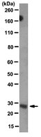
Evaluated by Western Blot in mouse brain microsomal tissue lysate.
Western Blot Analysis: 0.25 µg/mL of this antibody detected Neuregulin-1 on 10 µg of mouse brain microsomal tissue lysate.
Usage Statement
Unless otherwise stated in our catalog or other company documentation accompanying the product(s), our products are intended for research use only and are not to be used for any other purpose, which includes but is not limited to, unauthorized commercial uses, in vitro diagnostic uses, ex vivo or in vivo therapeutic uses or any type of consumption or application to humans or animals.
Neuregulin-1 modulates the differentiation of neural stem cells in vitro through an interaction with the Swi/Snf complex. Pirotte, D; Wislet-Gendebien, S; Cloes, JM; Rogister, B Molecular and cellular neurosciences
43
72-80
2009
The neuregulin-1 (Nrg-1) gene is translated into several protein isoforms, which are either secreted or membrane-anchored. In vitro, neural stem cells (NSC) express mainly the cystein-rich-domain NRG (CRD-NRG) isoform, a membrane-anchored type III form. This isoform exhibits a cystein-rich-domain, which constitutes a second transmembrane domain and can be cleaved to release both a signaling EGF-containing domain (ECD) at the cell surface and an intracellular domain (ICD). The main goal of this paper was to determine the exact role of ECD and ICD in NSC survival and differentiation. Using an siRNA approach, we demonstrated that CRD-NRG inhibition was followed by a decrease in NSC proliferation and of neuronal or oligodendroglial differentiation. Overexpression of ICD but not ECD was followed by a decrease in NSC proliferation and an increase in neuronal and oligodendroglial differentiation. Moreover, we showed that ICD physically interacted in cultured NSC with BRM and BAF57, two members of the Swi/Snf remodeling complex, and that ICD stimulation of neuronal cell differentiation is dependent on the presence of BAF57.