Wenn Sie das Fenster schließen, wird Ihre Konfiguration nicht gespeichert, es sei denn, Sie haben Ihren Artikel in die Bestellung aufgenommen oder zu Ihren Favoriten hinzugefügt.
Klicken Sie auf OK, um das MILLIPLEX® MAP-Tool zu schließen oder auf Abbrechen, um zu Ihrer Auswahl zurückzukehren.
Wählen Sie konfigurierbare Panels & Premixed-Kits - ODER - Kits für die zelluläre Signaltransduktion & MAPmates™
Konfigurieren Sie Ihre MILLIPLEX® MAP-Kits und lassen sich den Preis anzeigen.
Konfigurierbare Panels & Premixed-Kits
Unser breites Angebot enthält Multiplex-Panels, für die Sie die Analyten auswählen können, die am besten für Ihre Anwendung geeignet sind. Unter einem separaten Register können Sie das Premixed-Cytokin-Format oder ein Singleplex-Kit wählen.
Kits für die zelluläre Signaltransduktion & MAPmates™
Wählen Sie gebrauchsfertige Kits zur Erforschung gesamter Signalwege oder Prozesse. Oder konfigurieren Sie Ihre eigenen Kits mit Singleplex MAPmates™.
Die folgenden MAPmates™ sollten nicht zusammen analysiert werden: -MAPmates™, die einen unterschiedlichen Assaypuffer erfordern. -Phosphospezifische und MAPmate™ Gesamtkombinationen wie Gesamt-GSK3β und Gesamt-GSK3β (Ser 9). -PanTyr und locusspezifische MAPmates™, z.B. Phospho-EGF-Rezeptor und Phospho-STAT1 (Tyr701). -Mehr als 1 Phospho-MAPmate™ für ein einziges Target (Akt, STAT3). -GAPDH und β-Tubulin können nicht mit Kits oder MAPmates™, die panTyr enthalten, analysiert werden.
.
Bestellnummer
Bestellinformationen
St./Pkg.
Liste
Dieser Artikel wurde zu Ihren Favoriten hinzugefügt.
Wählen Sie bitte Spezies, Panelart, Kit oder Probenart
Um Ihr MILLIPLEX® MAP-Kit zu konfigurieren, wählen Sie zunächst eine Spezies, eine Panelart und/oder ein Kit.
Custom Premix Selecting "Custom Premix" option means that all of the beads you have chosen will be premixed in manufacturing before the kit is sent to you.
Catalogue Number
Ordering Description
Qty/Pack
List
Dieser Artikel wurde zu Ihren Favoriten hinzugefügt.
Spezies
Panelart
Gewähltes Kit
Menge
Bestellnummer
Bestellinformationen
St./Pkg.
Listenpreis
96-Well Plate
Menge
Bestellnummer
Bestellinformationen
St./Pkg.
Listenpreis
Weitere Reagenzien hinzufügen (MAPmates erfordern die Verwendung eines Puffer- und Detektionskits)
Menge
Bestellnummer
Bestellinformationen
St./Pkg.
Listenpreis
48-602MAG
Buffer Detection Kit for Magnetic Beads
1 Kit
Platzsparende Option Kunden, die mehrere Kits kaufen, können ihre Multiplex-Assaykomponenten in Kunststoffbeuteln anstelle von Packungen erhalten, um eine kompaktere Lagerung zu ermöglichen.
Dieser Artikel wurde zu Ihren Favoriten hinzugefügt.
Das Produkt wurde in Ihre Bestellung aufgenommen
Sie können nun ein weiteres Kit konfigurieren, ein Premixed-Kit wählen, zur Kasse gehen oder das Bestell-Tool schließen.
Detect Ras related protein Rab-10 using this mouse monoclonal antibody, Anti-Rab10 Antibody, clone 4E2 validated for use in western blotting & Immunofluorescence.
More>>Detect Ras related protein Rab-10 using this mouse monoclonal antibody, Anti-Rab10 Antibody, clone 4E2 validated for use in western blotting & Immunofluorescence. Less<<
Anti-Rab10 Antibody, clone 4E2: SDB (Sicherheitsdatenblätter), Analysenzertifikate und Qualitätszertifikate, Dossiers, Broschüren und andere verfügbare Dokumente.
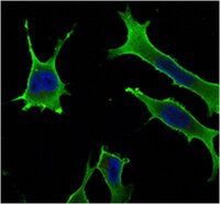
Ras related protein Rab-10 (Rab10) is encoded by the gene named RAB10 and is member of the small GTPase family of Rab proteins. Rab proteins play critical roles in intracellular trafficking from the formation of vesicles to their fusion with membranes. Specifically Rab10 is involved in regulating RME-1 in endocytic recycling as well as endoplasmic reticulum structural dynamics and morphology, and Rab10 also appears to be critical to formation and correct placement of proteins in such critical cellular structures as the basement membranes of during organ morphogenesis and basement membrane remodeling during development. Rab10 localizes to internal membrane structures like the endoplasmic reticulum and Golgi apparatus but in certain cells it can be found associated with cilia and other cellular structures as well. EMD-Millipore’s Anti-Rab10 mouse monoclonal antibody has been tested in western blot against human HeLa and mouse NIH3T3 cell lysates and in fluorescent immunocytochemistry on LOVO cells in culture.
References
Product Information
Format
Ascites
Control
HeLa and NIH/3T3 cell lysates
Presentation
Mouse monoclonal IgG1 ascitic fluid containing up to 0.1% sodium azide.
Detect Ras related protein Rab-10 using this mouse monoclonal antibody, Anti-Rab10 Antibody, clone 4E2 validated for use in western blotting & Immunofluorescence.
Key Applications
Western Blotting
Immunofluorescence
Application Notes
Immunofluorescence Analysis: A 1:200-1,000 dilution from a representative lot detected Rab10 in LOVO cells.
Optimal working dilutions must be determined by end user.
Biological Information
Immunogen
Purified recombinant fragment of human Rab10 expressed in E. Coli.
Clone
4E2
Host
Mouse
Isotype
IgG1
Species Reactivity
Human
Mouse
Antibody Type
Monoclonal Antibody
Gene Symbol
RAB10
Purification Method
Unpurified
Molecular Weight
~25 kDa observed. Uncharacterized bands may appear in some lysate(s).
Physicochemical Information
Dimensions
Materials Information
Toxicological Information
Safety Information according to GHS
Safety Information
Product Usage Statements
Quality Assurance
Evaluated by Western Blotting in HeLa and NIH/3T3 cell lysates.
Western Blotting Analysis: A 1:500-2,000 dilution of this antibody detected Rab10 in HeLa and NIH/3T3 cell lysates.
Usage Statement
Unless otherwise stated in our catalog or other company documentation accompanying the product(s), our products are intended for research use only and are not to be used for any other purpose, which includes but is not limited to, unauthorized commercial uses, in vitro diagnostic uses, ex vivo or in vivo therapeutic uses or any type of consumption or application to humans or animals.
Storage and Shipping Information
Storage Conditions
Stable for 1 year at -20°C from date of receipt. Handling Recommendations: Upon receipt and prior to removing the cap, centrifuge the vial and gently mix the solution. Aliquot into microcentrifuge tubes and store at -20°C. Avoid repeated freeze/thaw cycles, which may damage IgG and affect product performance.