Wenn Sie das Fenster schließen, wird Ihre Konfiguration nicht gespeichert, es sei denn, Sie haben Ihren Artikel in die Bestellung aufgenommen oder zu Ihren Favoriten hinzugefügt.
Klicken Sie auf OK, um das MILLIPLEX® MAP-Tool zu schließen oder auf Abbrechen, um zu Ihrer Auswahl zurückzukehren.
Wählen Sie konfigurierbare Panels & Premixed-Kits - ODER - Kits für die zelluläre Signaltransduktion & MAPmates™
Konfigurieren Sie Ihre MILLIPLEX® MAP-Kits und lassen sich den Preis anzeigen.
Konfigurierbare Panels & Premixed-Kits
Unser breites Angebot enthält Multiplex-Panels, für die Sie die Analyten auswählen können, die am besten für Ihre Anwendung geeignet sind. Unter einem separaten Register können Sie das Premixed-Cytokin-Format oder ein Singleplex-Kit wählen.
Kits für die zelluläre Signaltransduktion & MAPmates™
Wählen Sie gebrauchsfertige Kits zur Erforschung gesamter Signalwege oder Prozesse. Oder konfigurieren Sie Ihre eigenen Kits mit Singleplex MAPmates™.
Die folgenden MAPmates™ sollten nicht zusammen analysiert werden: -MAPmates™, die einen unterschiedlichen Assaypuffer erfordern. -Phosphospezifische und MAPmate™ Gesamtkombinationen wie Gesamt-GSK3β und Gesamt-GSK3β (Ser 9). -PanTyr und locusspezifische MAPmates™, z.B. Phospho-EGF-Rezeptor und Phospho-STAT1 (Tyr701). -Mehr als 1 Phospho-MAPmate™ für ein einziges Target (Akt, STAT3). -GAPDH und β-Tubulin können nicht mit Kits oder MAPmates™, die panTyr enthalten, analysiert werden.
.
Bestellnummer
Bestellinformationen
St./Pkg.
Liste
Dieser Artikel wurde zu Ihren Favoriten hinzugefügt.
Wählen Sie bitte Spezies, Panelart, Kit oder Probenart
Um Ihr MILLIPLEX® MAP-Kit zu konfigurieren, wählen Sie zunächst eine Spezies, eine Panelart und/oder ein Kit.
Custom Premix Selecting "Custom Premix" option means that all of the beads you have chosen will be premixed in manufacturing before the kit is sent to you.
Catalogue Number
Ordering Description
Qty/Pack
List
Dieser Artikel wurde zu Ihren Favoriten hinzugefügt.
Spezies
Panelart
Gewähltes Kit
Menge
Bestellnummer
Bestellinformationen
St./Pkg.
Listenpreis
96-Well Plate
Menge
Bestellnummer
Bestellinformationen
St./Pkg.
Listenpreis
Weitere Reagenzien hinzufügen (MAPmates erfordern die Verwendung eines Puffer- und Detektionskits)
Menge
Bestellnummer
Bestellinformationen
St./Pkg.
Listenpreis
48-602MAG
Buffer Detection Kit for Magnetic Beads
1 Kit
Platzsparende Option Kunden, die mehrere Kits kaufen, können ihre Multiplex-Assaykomponenten in Kunststoffbeuteln anstelle von Packungen erhalten, um eine kompaktere Lagerung zu ermöglichen.
Dieser Artikel wurde zu Ihren Favoriten hinzugefügt.
Das Produkt wurde in Ihre Bestellung aufgenommen
Sie können nun ein weiteres Kit konfigurieren, ein Premixed-Kit wählen, zur Kasse gehen oder das Bestell-Tool schließen.
Anti-CD69, clone FN61, Cat. No. MABF2210, is a mouse monoclonal antibody that detects Early activation antigen CD69 and has been tested for use in Combined Interference Assay, Flow Cytometry, Function Analysis, and Immunofluorescence.
More>>Anti-CD69, clone FN61, Cat. No. MABF2210, is a mouse monoclonal antibody that detects Early activation antigen CD69 and has been tested for use in Combined Interference Assay, Flow Cytometry, Function Analysis, and Immunofluorescence. Less<<
Empfohlene Produkte
Übersicht
Replacement Information
Description
Catalogue Number
MABF2210-100UG
Description
Anti-CD69 Antibody, clone FN61
Alternate Names
Early activation antigen CD69
Activation inducer molecule
AIM
BL-AC/P26
C-type lectin domain family 2 member C
EA1
Early T-cell activation antigen p60
GP32/28
Leukocyte surface antigen Leu-23
MLR-3
Background Information
Early activation antigen CD69 (UniProt: Q07108; also known as Activation inducer molecule, AIM, BL-AC/P26, C-type lectin domain family 2 member C, EA1, Early T-cell activation antigen p60, GP32/28, Leukocyte surface antigen Leu-23, MLR-3, CD69) is encoded by the CD69 (also known as CLEC2C) gene (Gene ID: 969) in human. CD69 is a single-pass type II membrane disulfide-linked homodimeric protein. It is a C-type lectin and a member of the natural killer (NK) receptor family and its expression is readily upregulated upon activation in most leukocytes. It is induced by antigens, mitogens or activators of protein kinase C on the surface of T and B-lymphocytes and NK cells. It is shown to be involved in immune cell homeostasis, regulating the T cell-mediated immune response through the control of Th17 cell differentiation. Expression of CD69 by activated T lymphocytes is reported to trigger an anti-inflammatory mechanism mediated by Galectin-1, which regulates the immune response and prevents pathogenic Th17 responses. CD69 knockout mice are shown to develop an exacerbated form of collagen-induced arthritis. CD69 has a cytoplasmic domain (aa 1-40), a short transmembrane domain (aa 41-61), and an extracellular domain (aa 62-199). (Ref.: Sanchez-Mateos, P., and Sanchez-Madrid, F. (1991). Eur. J. Immunol. 21: 2317-2325; Ziegler SF et al., (1994). Stem Cells. 12(5); 456-65; de la Fuente, H., et al. (2014). Mol. Cell. Biol. 34(13); 2479-2487).
References
Product Information
Format
Purified
Presentation
Purified mouse monoclonal antibody IgG1 in PBS without azide.
Applications
Application
Anti-CD69, clone FN61, Cat. No. MABF2210, is a mouse monoclonal antibody that detects Early activation antigen CD69 and has been tested for use in Combined Interference Assay, Flow Cytometry, Function Analysis, and Immunofluorescence.
Combined Interference Assay: A representative lot was found to be suitable for combined interference assay. (Frengen, J., et. al. (1994). Clin Chem. 40(3):420-5).
Immunofluorescence Analysis: A representative lot detected CD69 in Immunofluorescence applications (Farstad, I.N., et. al. (1994). Immunology. 83(3):457-64).
Function Analysis: A representative lot triggered a strong proliferation in T lymphocytes. (Sanchez-Mateos, P., et. al. (1991). Eur J Immunol. 21(10):2317-25).
Biological Information
Immunogen
Activated B cells from human peripheral blood.
Epitope
extracellular domain
Clone
FN61
Concentration
Please refer to lot specific datasheet.
Host
Mouse
Specificity
Clone FN61 is a mouse monoclonal antibody that detects human Early activation antigen CD69. It targets an epitope within the E1 site in the extracellular domain.
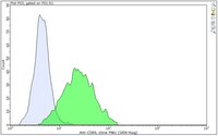
Evaluated by Flow Cytometry in Raji cells pretreated with 50 ng/mL PMA and 1 ug/mL ionomycin.
Flow Cytometry Analysis: 1 µg of this antibody detected CD69 in one million Raji cells pretreated with Phorbol 12-myristate 13-acetate (PMA; 50 ng/mL) and Ionomycin (1 ug/mL) for 4 to 6 hours.
Usage Statement
Unless otherwise stated in our catalog or other company documentation accompanying the product(s), our products are intended for research use only and are not to be used for any other purpose, which includes but is not limited to, unauthorized commercial uses, in vitro diagnostic uses, ex vivo or in vivo therapeutic uses or any type of consumption or application to humans or animals.
Storage and Shipping Information
Storage Conditions
Stable for 1 year at -20°C from date of receipt. Handling Recommendations: Upon receipt and prior to removing the cap, centrifuge the vial and gently mix the solution. Aliquot into microcentrifuge tubes and store at -20°C. Avoid repeated freeze/thaw cycles, which may damage IgG and affect product performance.
The ability of serum factors to cross-link labeled mouse monoclonal antibody (mAb) of irrelevant specificity (mAb FN61, subclass IgG1) to different particle types coated with sheep IgG, bovine gamma-globulin, or mAb FN61 was measured simultaneously by flow cytometry. Significant interference with mAb FN61-coated particles was detected in 53 of 101 sera. Of the 30 sera showing the most pronounced interference, 23 were characterized by an even stronger cross-linking to particles coated with bovine gamma-globulin. These were designated type 1 sera. Seven sera, designated type 2, displayed a dominant interference with the mAb FN61-coated particles. The interference reaction in the two serum types was characterized by different kinetics, dependence on particle concentration, and response to blocking agents. The interference was minimized by addition of 500 micrograms of bovine gamma-globulin and 50 micrograms of mAb HH1 (IgG1) of irrelevant specificity per 10 microL of serum sample in a final assay volume of 100 microL.
The CD53 pan-leukocyte glycoprotein is a member of the recently described tetraspan family of cell membrane proteins. The structure and functional characteristics of these molecules indicate that they may play important roles in transmembrane signaling in different cells. Recently, it was reported that cross-linking of CD53 on human B cells led to an increase in cytoplasmic calcium fluxes. In the present study, we wished to further explore the possible role of CD53 in functional B cell responses. Cross-linking of CD53 with the use of the mAb MEM-53 and a polyclonal sheep anti-mouse Ig promoted activation of resting B cells into the G1 phase of the cell cycle as judged by increased expression of the early activation Ag CD69, increases in cellular volume, RNA synthesis, and c-myc protein levels, and enhanced binding of 7-aminoactinomycin D. In contrast, MEM-53 alone had no detectable effects. Cross-linking of anti-CD53 induced negligible S phase entry in the absence of other stimuli. However, cytokines, in particular IL-2 and IL-4, potentiated the DNA synthesis induced by cross-linking of CD53. Furthermore, cross-linking of the CD53 Ag induced Ig production in the presence of T cell supernatant. Taken together, the data suggest that CD53 plays an important functional role in B cell activation and differentiation.
Heterogeneity of M-cell-associated B and T cells in human Peyer's patches. Farstad, IN; Halstensen, TS; Fausa, O; Brandtzaeg, P Immunology
83
457-64
1993
The specialized M cells in the follicle-associated epithelium (FAE) of Peyer's patches (PP) represent an intimate interphase between luminal antigens and gut-associated lymphoid tissue (GALT). M cells form pockets that contain clusters of leucocytes probably involved in the first encounter with antigens from the gut lumen. Three-colour immunofluorescence in situ phenotyping of these leucocytes in humans revealed about equal numbers of B (CD19/20+) and T(CD3+) lymphocytes, the latter mainly CD4+ (median 73%, range 40-90%), but relatively few macrophages (CD68+). Most B cells (90%) were positive for surface IgM (sIgM) and often co-expressed sIgD (median 34%, range 6-60%). Occasional B cells (median 2%) did not express CD45RA (range 0-15%) and 13% virtually lacked HLA-DR (range 0-40%). Some B and T lymphocytes expressed the nuclear proliferation marker Ki-67 (range 1-10%). The M-cell pockets also contained occasional cells with cytoplasmic IgA or IgM. These sites thus contained a heterogeneous B-cell population with features of both follicular mantle (sIgD+ sIgM+) and marginal zone (sIgD- sIgM+) B lymphocytes. Adjacent T lymphocytes were generally of the memory phenotype (CD45RO+). Our findings suggest that the M-cell-associated B lymphocytes represent local extensions of B-cell follicles towards the gut lumen, developed topically to facilitate antigen presentation and diversification of mucosal immune responses.
We evaluated two homogeneous immunofluorometric assays (IFMAs) of alpha-fetoprotein (AFP) based on new macroporous acrylate particles combined with flow cytometry. The standard IFMA, requiring 1 h of incubation, provided a working range from 1.8 to > 900 kIU/L (CV < 10%) and a detection limit of 0.6 kIU/L. Use of overnight incubation and a lower particle concentration extended the working range by 1 decade in the lower end. Analytical recoveries for the standard IFMA varied between 97% and 108%. The slope and y-intercept of the regression line correlating measurements by the standard IFMA and a routine immunoradiometric assay were not significantly different from 1 and 0, respectively (P > 0.5), and the correlation coefficient was 0.996. High precision and warning of spuriously high measurements were obtained by including in each sample separate particle types for detecting instrument instability and measuring nonspecific binding only.
Structure-function relationship and immunochemical mapping of external and intracellular antigenic sites on the lymphocyte activation inducer molecule, AIM/CD69. Sánchez-Mateos, P; Sánchez-Madrid, F Eur J Immunol
21
2317-25
1991
Human activation inducer molecule (AIM/CD69), a dimeric glycoprotein structure of 33 and 27 kDa, is the earliest inducible cell surface antigen expressed during lymphocyte activation and is implicated in the induction of T and B cell proliferative responses. Cross-competition monoclonal antibodies (mAb) binding assays have allowed the definition of four antigenic epitopes. Three of them (antigenic sites E1-3) are extracellular while the fourth (site I) is a sequential epitope localized intracellularly and highly conserved interspecies. Site E1 is shown to be an immunodominant antigenic determinant closely related to a functional domain of AIM important for triggering of T cell proliferation. Studies of peptide fragmentation of the two isolated AIM subunits with different proteases have demonstrated that both AIM chains are differentially glycosylated forms of a single 24-kDa core protein. Moreover, the two denatured and isolated AIM chains share common epitope(s) as demonstrated by their reactivity with an mAb by both Western blot analysis and immunoprecipitation of the separated AIM subunits. Biosynthesis studies revealed the rapid appearance of two intermediate precursor forms of 29 and 26 kDa which arise from the 24-kDa unglycosylated AIM polypeptide. This 24-kDa unglycosylated form could be also precipitated from iodinated cells pretreated with tunicamycin, indicating that glycosylation of the protein was neither required for AIM cell surface expression nor for acquisition of external epitopes E1-E3. Cell treatment with pronase resulted in the loss of the external epitopes E1-3 and the generation of a proteolytic peptide of 16 kDa that could be precipitated by the anti-AIM mAb specific for the internal site I. This proteolytic fragment retained the transmembrane and cytoplasmic regions of the molecule where both epitope I and phosphorylation sites reside. These results demonstrate that AIM is an integral membrane homodimeric glycoprotein with a large cytoplasmic domain probably involved in the activation signals transduced through this molecule to lymphocytes.