Wenn Sie das Fenster schließen, wird Ihre Konfiguration nicht gespeichert, es sei denn, Sie haben Ihren Artikel in die Bestellung aufgenommen oder zu Ihren Favoriten hinzugefügt.
Klicken Sie auf OK, um das MILLIPLEX® MAP-Tool zu schließen oder auf Abbrechen, um zu Ihrer Auswahl zurückzukehren.
Wählen Sie konfigurierbare Panels & Premixed-Kits - ODER - Kits für die zelluläre Signaltransduktion & MAPmates™
Konfigurieren Sie Ihre MILLIPLEX® MAP-Kits und lassen sich den Preis anzeigen.
Konfigurierbare Panels & Premixed-Kits
Unser breites Angebot enthält Multiplex-Panels, für die Sie die Analyten auswählen können, die am besten für Ihre Anwendung geeignet sind. Unter einem separaten Register können Sie das Premixed-Cytokin-Format oder ein Singleplex-Kit wählen.
Kits für die zelluläre Signaltransduktion & MAPmates™
Wählen Sie gebrauchsfertige Kits zur Erforschung gesamter Signalwege oder Prozesse. Oder konfigurieren Sie Ihre eigenen Kits mit Singleplex MAPmates™.
Die folgenden MAPmates™ sollten nicht zusammen analysiert werden: -MAPmates™, die einen unterschiedlichen Assaypuffer erfordern. -Phosphospezifische und MAPmate™ Gesamtkombinationen wie Gesamt-GSK3β und Gesamt-GSK3β (Ser 9). -PanTyr und locusspezifische MAPmates™, z.B. Phospho-EGF-Rezeptor und Phospho-STAT1 (Tyr701). -Mehr als 1 Phospho-MAPmate™ für ein einziges Target (Akt, STAT3). -GAPDH und β-Tubulin können nicht mit Kits oder MAPmates™, die panTyr enthalten, analysiert werden.
.
Bestellnummer
Bestellinformationen
St./Pkg.
Liste
Dieser Artikel wurde zu Ihren Favoriten hinzugefügt.
Wählen Sie bitte Spezies, Panelart, Kit oder Probenart
Um Ihr MILLIPLEX® MAP-Kit zu konfigurieren, wählen Sie zunächst eine Spezies, eine Panelart und/oder ein Kit.
Custom Premix Selecting "Custom Premix" option means that all of the beads you have chosen will be premixed in manufacturing before the kit is sent to you.
Catalogue Number
Ordering Description
Qty/Pack
List
Dieser Artikel wurde zu Ihren Favoriten hinzugefügt.
Spezies
Panelart
Gewähltes Kit
Menge
Bestellnummer
Bestellinformationen
St./Pkg.
Listenpreis
96-Well Plate
Menge
Bestellnummer
Bestellinformationen
St./Pkg.
Listenpreis
Weitere Reagenzien hinzufügen (MAPmates erfordern die Verwendung eines Puffer- und Detektionskits)
Menge
Bestellnummer
Bestellinformationen
St./Pkg.
Listenpreis
48-602MAG
Buffer Detection Kit for Magnetic Beads
1 Kit
Platzsparende Option Kunden, die mehrere Kits kaufen, können ihre Multiplex-Assaykomponenten in Kunststoffbeuteln anstelle von Packungen erhalten, um eine kompaktere Lagerung zu ermöglichen.
Dieser Artikel wurde zu Ihren Favoriten hinzugefügt.
Das Produkt wurde in Ihre Bestellung aufgenommen
Sie können nun ein weiteres Kit konfigurieren, ein Premixed-Kit wählen, zur Kasse gehen oder das Bestell-Tool schließen.
ABT119
Sigma-AldrichAnti-CELSR1 Antibody
This Anti-CELSR1 Antibody is validated for use in Western Blotting, IHC, Immunofluorescence for the detection of CELSR1.
More>>This Anti-CELSR1 Antibody is validated for use in Western Blotting, IHC, Immunofluorescence for the detection of CELSR1. Less<<
Anti-CELSR1 Antibody: SDB (Sicherheitsdatenblätter), Analysenzertifikate und Qualitätszertifikate, Dossiers, Broschüren und andere verfügbare Dokumente.
The Flamingo/CELSR family of 7TM-cadherins are characterized with a symmetric protein distribution and polarized activity at neighboring epithelial cell interfaces along defined axes of planar cell polarity. As a component of the extracellular matrix, the CELSR1 protein has been found particularly at the basal surface of neuroepithelial cells within both the early neural tube and a less well-defined group of ventricular zone cells at the midline of the developing spinal cord. CELSR1 plays a role in orienting hair follicles along the anterior–posterior axis of the developing mouse epidermis, both during embryogenesis and postnatal development.
References
Product Information
Format
Affinity Purified
Control
PC12 cell lysate.
Presentation
Purified rabbit polyclonal in buffer containing 0.1 M Tris-Glycine (pH 7.4), 150 mM NaCl with 0.05% sodium azide.
This Anti-CELSR1 Antibody is validated for use in Western Blotting, IHC, Immunofluorescence for the detection of CELSR1.
Key Applications
Western Blotting
Immunohistochemistry
Immunofluorescence
Application Notes
Immunofluorescence Analysis: A 1:500 and 1:1,000 dilution from a representative lot detected CELSR1 in rat cerebellum, substancia nigra, and hippocampus tissue.
Immunohistochemistry Analysis: A 1:500 dilution from a representative lot detected CELSR1 in neurons of rat cortex tissue and in Purkinje cells and cells within the granular layer of rat cerebellum tissue.
Biological Information
Immunogen
Linear peptide corresponding to human CELSR1.
Concentration
Please refer to the Certificate of Analysis for the lot-specific concentration.
Host
Rabbit
Species Reactivity
Rat
Human
Mouse
Species Reactivity Note
Demonstrated to react with Rat. Predicted to react with Human and Mouse based on 100% sequence homology.
The protein encoded by this gene is a member of the flamingo subfamily, part of the cadherin superfamily. The flamingo subfamily consists of nonclassic-type cadherins; a subpopulation that does not interact with catenins. The flamingo cadherins are located at the plasma membrane and have nine cadherin domains, seven epidermal growth factor-like repeats and two laminin A G-type repeats in their ectodomain. They also have seven transmembrane domains, a characteristic unique to this subfamily. It is postulated that these proteins are receptors involved in contact-mediated communication, with cadherin domains acting as homophilic binding regions and the EGF-like domains involved in cell adhesion and receptor-ligand interactions. This particular member is a developmentally regulated, neural-specific gene which plays an unspecified role in early embryogenesis.
POST-TRANSLATIONAL MODIFICATION: The iron and 2-oxoglutarate dependent 3-hydroxylation of aspartate and asparagine is (R) stereospecific within EGF domains.
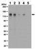
~330 kDa observed. A second isoform at ~152 kDa may be observed in some lysates. An uncharacterized band at ~54 kDa may be observed in some cell lysates.
Physicochemical Information
Dimensions
Materials Information
Toxicological Information
Safety Information according to GHS
Safety Information
Product Usage Statements
Quality Assurance
Evaluated by Western Blot in PC12 cell lysate.
Western Blot Analysis: 1 µg/mL of this antibody detected CELSR1 in 10 µg of PC12 cell lysate.
Usage Statement
Unless otherwise stated in our catalog or other company documentation accompanying the product(s), our products are intended for research use only and are not to be used for any other purpose, which includes but is not limited to, unauthorized commercial uses, in vitro diagnostic uses, ex vivo or in vivo therapeutic uses or any type of consumption or application to humans or animals.