Wenn Sie das Fenster schließen, wird Ihre Konfiguration nicht gespeichert, es sei denn, Sie haben Ihren Artikel in die Bestellung aufgenommen oder zu Ihren Favoriten hinzugefügt.
Klicken Sie auf OK, um das MILLIPLEX® MAP-Tool zu schließen oder auf Abbrechen, um zu Ihrer Auswahl zurückzukehren.
Wählen Sie konfigurierbare Panels & Premixed-Kits - ODER - Kits für die zelluläre Signaltransduktion & MAPmates™
Konfigurieren Sie Ihre MILLIPLEX® MAP-Kits und lassen sich den Preis anzeigen.
Konfigurierbare Panels & Premixed-Kits
Unser breites Angebot enthält Multiplex-Panels, für die Sie die Analyten auswählen können, die am besten für Ihre Anwendung geeignet sind. Unter einem separaten Register können Sie das Premixed-Cytokin-Format oder ein Singleplex-Kit wählen.
Kits für die zelluläre Signaltransduktion & MAPmates™
Wählen Sie gebrauchsfertige Kits zur Erforschung gesamter Signalwege oder Prozesse. Oder konfigurieren Sie Ihre eigenen Kits mit Singleplex MAPmates™.
Die folgenden MAPmates™ sollten nicht zusammen analysiert werden: -MAPmates™, die einen unterschiedlichen Assaypuffer erfordern. -Phosphospezifische und MAPmate™ Gesamtkombinationen wie Gesamt-GSK3β und Gesamt-GSK3β (Ser 9). -PanTyr und locusspezifische MAPmates™, z.B. Phospho-EGF-Rezeptor und Phospho-STAT1 (Tyr701). -Mehr als 1 Phospho-MAPmate™ für ein einziges Target (Akt, STAT3). -GAPDH und β-Tubulin können nicht mit Kits oder MAPmates™, die panTyr enthalten, analysiert werden.
.
Bestellnummer
Bestellinformationen
St./Pkg.
Liste
Dieser Artikel wurde zu Ihren Favoriten hinzugefügt.
Wählen Sie bitte Spezies, Panelart, Kit oder Probenart
Um Ihr MILLIPLEX® MAP-Kit zu konfigurieren, wählen Sie zunächst eine Spezies, eine Panelart und/oder ein Kit.
Custom Premix Selecting "Custom Premix" option means that all of the beads you have chosen will be premixed in manufacturing before the kit is sent to you.
Catalogue Number
Ordering Description
Qty/Pack
List
Dieser Artikel wurde zu Ihren Favoriten hinzugefügt.
Spezies
Panelart
Gewähltes Kit
Menge
Bestellnummer
Bestellinformationen
St./Pkg.
Listenpreis
96-Well Plate
Menge
Bestellnummer
Bestellinformationen
St./Pkg.
Listenpreis
Weitere Reagenzien hinzufügen (MAPmates erfordern die Verwendung eines Puffer- und Detektionskits)
Menge
Bestellnummer
Bestellinformationen
St./Pkg.
Listenpreis
48-602MAG
Buffer Detection Kit for Magnetic Beads
1 Kit
Platzsparende Option Kunden, die mehrere Kits kaufen, können ihre Multiplex-Assaykomponenten in Kunststoffbeuteln anstelle von Packungen erhalten, um eine kompaktere Lagerung zu ermöglichen.
Dieser Artikel wurde zu Ihren Favoriten hinzugefügt.
Das Produkt wurde in Ihre Bestellung aufgenommen
Sie können nun ein weiteres Kit konfigurieren, ein Premixed-Kit wählen, zur Kasse gehen oder das Bestell-Tool schließen.
Anti-Hypoxia Inducible Factor 2a , clone 190b, Cat. No. MAB3472-I, is a mouse monoclonal antibody that detects Hypoxia Inducible Factor 2a and is tested for use in Immunocytochemistry, Immunohistochemistry (Paraffin), and Western Blotting.
More>>Anti-Hypoxia Inducible Factor 2a , clone 190b, Cat. No. MAB3472-I, is a mouse monoclonal antibody that detects Hypoxia Inducible Factor 2a and is tested for use in Immunocytochemistry, Immunohistochemistry (Paraffin), and Western Blotting. Less<<
Endothelial PAS domain-containing protein 1 (UniProt: Q99814; also known as EPAS-1, Basic-helix-loop-helix-PAS protein MOP2, Class E basic helix-loop-helix protein 73, bHLHe73, HIF-1-alpha-like factor, HLF, Hypoxia-inducible factor 2-alpha, HIF-2-alpha, HIF2-alpha, Member of PAS protein 2, PAS domain-containing protein 2) is encoded by the EPAS1 (also known as BHLHE73, HIF2A, MOP2, PASD2) gene (Gene ID: 2034) in human. HIF-2a is a transcription factor that is involved in the induction of oxygen regulated genes. It displays about 48% identity to HIF-1a and is widely expressed with high expression observed in endothelial cells. Its levels are strongly induced by hypoxic conditions. HIF- 2a dimerizes with ARNT, via amino acid sequence 171-192, and this heterodimer binds to core DNA sequence 5'-TACGTG-3' within the hypoxia response element (HRE) of target gene promoters. Its DNA-binding region is localized to amino acids 26-53. It has two PAS (Per-Arnt-Sim) domains (aa 84-154 and 23-300). HIF-2a regulates the vascular endothelial growth factor (VEGF) expression and is implicated in the development of blood vessels and the tubular system of lung and is also involved in the formation of the endothelium that gives rise to the blood brain barrier. Under normoxic conditions it is hydroxylated on proline 405 and 531 by prolyl hydroxylase 1 and 2 and the hydroxylated form undergoes rapid ubiquitination and proteasomal degradation. However, under hypoxic conditions, proline hydroxylation is impaired and HIF- 2a is stabilized. Mutations in EPAS1 gene are known to cause familial type erythrocytosis that is characterized by elevated serum hemoglobin and hematocrit with normal platelet and leukocyte counts. (Ref.: Strowitzki, MJ., et al. (2019). Cells. 8(5): 384; Wiesener, MS., et al. (1998). Blood. 92(7); 2260-2268).
References
Product Information
Format
Purified
Presentation
Purified mouse monoclonal antibody IgG1 in buffer containing 0.1 M Tris-Glycine (pH 7.4), 150 mM NaCl with 0.05% sodium azide.
Anti-Hypoxia Inducible Factor 2a , clone 190b, Cat. No. MAB3472-I, is a mouse monoclonal antibody that detects Hypoxia Inducible Factor 2a and is tested for use in Immunocytochemistry, Immunohistochemistry (Paraffin), and Western Blotting.
Key Applications
Immunocytochemistry
Immunohistochemistry (Paraffin)
Western Blotting
Application Notes
Tested Applications
Immunohistochemistry Applications: A representative lot detected Hypoxia Inducible Factor 2 in Immunohistochemistry applications (Hoefflin, R. et al. (2020). Nat Commun.;11(1):4111; Moriyama, H., et al. (2014). Stem Cells Dev.;23(18):2211-24).
Immunohistochemistry (Paraffin) Analysis: A 1:50 dilution from a representative lot detected Hypoxia Inducible Factor 2 in human bone marrow tissue sections.
Immunocytochemistry Analysis: A representative lot detected Hypoxia Inducible Factor 2 in Immunocytochemistry applications (Talks, K.L., et al. (2000). Am J Pathol.;157(2):411-21; Wiesener, M.S., et al. (1998). Blood.;92(7):2260-8).
Western Blotting Analysis: A representative lot detected Hypoxia Inducible Factor 2 in Western Blotting applications (Ma, Y., et al. (2015). EMBO J.;34(3):361-78; Talks, K.L., et al. (2000). Am J Pathol.;157(2):411-21; Wiesener, M.S., et al. (1998). Blood.;92(7):2260-8).
Immunohistochemistry (Paraffin) Analysis: A representative lot detected Hypoxia Inducible Factor 2 in Immunohistochemistry applications (Talks, K.L., et al. (2000). Am J Pathol.;157(2):411-21).
Note: Actual optimal working dilutions must be determined by end user as specimens, and experimental conditions may vary with the end user
Biological Information
Immunogen
GST-tagged recombinant fragment corresponding to 97 amino acids from the C-terminal half of human Hypoxia inducible factor 2a (HIF- 2a).
Epitope
C-terminal half
Clone
190b
Concentration
0.5 mg/mL. Please refer to guidance on suggested starting dilutions and/or titers per application and sample type.
Host
Mouse
Specificity
Clone 190b is a mouse monoclonal antibody that detects Hypoxia-inducible factor 2a. It targets an epitope within the C-terminal half.
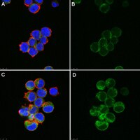
Evaluated by Immunocytochemistry in hypoxic Jurkat cells.
Immunocytochemistry Analysis (ICC): A 1:500 dilution of this antibody detected HIF-2a in hypoxic Jurkat cells.
Usage Statement
Unless otherwise stated in our catalog or other company documentation accompanying the product(s), our products are intended for research use only and are not to be used for any other purpose, which includes but is not limited to, unauthorized commercial uses, in vitro diagnostic uses, ex vivo or in vivo therapeutic uses or any type of consumption or application to humans or animals.
Storage and Shipping Information
Storage Conditions
Recommend storage at +2°C to +8°C. For long term storage antibodies can be kept at -20°C. Avoid repeated freeze-thaws.