Wenn Sie das Fenster schließen, wird Ihre Konfiguration nicht gespeichert, es sei denn, Sie haben Ihren Artikel in die Bestellung aufgenommen oder zu Ihren Favoriten hinzugefügt.
Klicken Sie auf OK, um das MILLIPLEX® MAP-Tool zu schließen oder auf Abbrechen, um zu Ihrer Auswahl zurückzukehren.
Wählen Sie konfigurierbare Panels & Premixed-Kits - ODER - Kits für die zelluläre Signaltransduktion & MAPmates™
Konfigurieren Sie Ihre MILLIPLEX® MAP-Kits und lassen sich den Preis anzeigen.
Konfigurierbare Panels & Premixed-Kits
Unser breites Angebot enthält Multiplex-Panels, für die Sie die Analyten auswählen können, die am besten für Ihre Anwendung geeignet sind. Unter einem separaten Register können Sie das Premixed-Cytokin-Format oder ein Singleplex-Kit wählen.
Kits für die zelluläre Signaltransduktion & MAPmates™
Wählen Sie gebrauchsfertige Kits zur Erforschung gesamter Signalwege oder Prozesse. Oder konfigurieren Sie Ihre eigenen Kits mit Singleplex MAPmates™.
Die folgenden MAPmates™ sollten nicht zusammen analysiert werden: -MAPmates™, die einen unterschiedlichen Assaypuffer erfordern. -Phosphospezifische und MAPmate™ Gesamtkombinationen wie Gesamt-GSK3β und Gesamt-GSK3β (Ser 9). -PanTyr und locusspezifische MAPmates™, z.B. Phospho-EGF-Rezeptor und Phospho-STAT1 (Tyr701). -Mehr als 1 Phospho-MAPmate™ für ein einziges Target (Akt, STAT3). -GAPDH und β-Tubulin können nicht mit Kits oder MAPmates™, die panTyr enthalten, analysiert werden.
.
Bestellnummer
Bestellinformationen
St./Pkg.
Liste
Dieser Artikel wurde zu Ihren Favoriten hinzugefügt.
Wählen Sie bitte Spezies, Panelart, Kit oder Probenart
Um Ihr MILLIPLEX® MAP-Kit zu konfigurieren, wählen Sie zunächst eine Spezies, eine Panelart und/oder ein Kit.
Custom Premix Selecting "Custom Premix" option means that all of the beads you have chosen will be premixed in manufacturing before the kit is sent to you.
Catalogue Number
Ordering Description
Qty/Pack
List
Dieser Artikel wurde zu Ihren Favoriten hinzugefügt.
Spezies
Panelart
Gewähltes Kit
Menge
Bestellnummer
Bestellinformationen
St./Pkg.
Listenpreis
96-Well Plate
Menge
Bestellnummer
Bestellinformationen
St./Pkg.
Listenpreis
Weitere Reagenzien hinzufügen (MAPmates erfordern die Verwendung eines Puffer- und Detektionskits)
Menge
Bestellnummer
Bestellinformationen
St./Pkg.
Listenpreis
48-602MAG
Buffer Detection Kit for Magnetic Beads
1 Kit
Platzsparende Option Kunden, die mehrere Kits kaufen, können ihre Multiplex-Assaykomponenten in Kunststoffbeuteln anstelle von Packungen erhalten, um eine kompaktere Lagerung zu ermöglichen.
Dieser Artikel wurde zu Ihren Favoriten hinzugefügt.
Das Produkt wurde in Ihre Bestellung aufgenommen
Sie können nun ein weiteres Kit konfigurieren, ein Premixed-Kit wählen, zur Kasse gehen oder das Bestell-Tool schließen.
This hamster monoclonal Anti-Neuroblasts Antibody, clone Nilo2, Cat. No. MABD402 is validated for use in Flow Cytometry, Immunocytochemistry, Immunohistochemistry, Immunofluorescence, Immunoprecipitation, Magnetic Resonance Imaging, Inhibition, and Western Blotting.
More>>This hamster monoclonal Anti-Neuroblasts Antibody, clone Nilo2, Cat. No. MABD402 is validated for use in Flow Cytometry, Immunocytochemistry, Immunohistochemistry, Immunofluorescence, Immunoprecipitation, Magnetic Resonance Imaging, Inhibition, and Western Blotting. Less<<
Anti-Neuroblasts Antibody, clone Nilo2: SDB (Sicherheitsdatenblätter), Analysenzertifikate und Qualitätszertifikate, Dossiers, Broschüren und andere verfügbare Dokumente.
FC, IHC, IP, Inhibition, Magnetic Resonance Imaging, WB
AHm
Purified
Monoclonal Antibody
Description
Catalogue Number
MABD402
Description
Anti-Neuroblasts Antibody, clone Nilo2
Alternate Names
Neuroblasts
Neuronal progenitors
Type A cells
Background Information
Similar to other cell types, neural cells are generated from primary progenitor cells known as type B astrocytes or neural stem cells (NSCs) via stages of amplification (transient amplifying cells), resulting in the generation of intermediate precursor cells (iPCs) committed to neurons (nIPCs), astrocytes (aIPCs), or oligodendrocytes (oIPCs). In adult rodent brain, NSCs and iPCs are mainly restricted to niches in the subventricular zone (SVZ) of the lateral ventricles (LV). In the adult SVZ, type B cells (SVZB) generate neuroblasts (type A cells, neuronal precursors) through a highly proliferative transit amplifying population (type C cells). NSCs and iPCs paly important roles in neurogenesis in the adult brain. Neuroblasts, for example, are known to migrate from their SVZ niche through the rostral migratory stream (RMS) to the olfactory bulb for the continuous generation of periglomerular interneurons. In addition to maintaining the homeostasis of the olfactory bulb, both neuroblasts and NSCs are shown to mobilize from their SVZ niches to various brain damage sites caused by tumor growth, cryolesion, focal demyelinization, and mechanical lesion.
References
Product Information
Format
Purified
Presentation
Purified Armenian hamster monoclonal antibody in PBS without preservatives.
This hamster monoclonal Anti-Neuroblasts Antibody, clone Nilo2, Cat. No. MABD402 is validated for use in Flow Cytometry, Immunocytochemistry, Immunohistochemistry, Immunofluorescence, Immunoprecipitation, Magnetic Resonance Imaging, Inhibition, and Western Blotting.
Key Applications
Flow Cytometry
Immunohistochemistry
Immunoprecipitation
Inhibition
Magnetic Resonance Imaging
Western Blotting
Application Notes
Magnetic Resonance Imaging (MRI) Analysis: A representative lot, when coupled to magnetic glyconanoparticles (mGNPs) and injected in mice, allowed the tracking of labelled neuroblasts migrating from their niches towards brain injury sites by MRI (Elvira, G., et al. (2015). Stem Cell Res. 14(1):114-129; Elvira, G., et al. (2012). PLoS One. 7(9):e44466).
Flow Cytometry Analysis: A representative lot, coupled to magnetic glyconanoparticles (mGNPs) or not, immunostained the surface of SVZ neurosphere-derived single cells (Elvira, G., et al. (2012). PLoS One. 7(9):e44466).
Flow Cytometry Analysis: Clone Nilo2 hybridoma culture supernatant stained the surface of 98% and 95% neurosphere-derived cells from mouse embryo olfactory bulb (OB) and adult mouse subventricular zone (SVZ), respectively (Del Valle, I., et al. (2010). Neuroscience. 169(3):1473-1485).
Immunocytochemistry Analysis: Representative lot immunostained early neural precursor cells by fluorescent immunocytochemistry staining of cultured neurospheres or neurosphere-derived single cells fixed with 4% paraformaldehyde. Unlike Nilo1 (Cat. No. MABD401), a small percentage of cells retained Nilo2 immunoreactivity even 7 days after differentiation induction by EGF and bFGF withdrawal in neurosphere cultures (Elvira, G., et al. (2012). PLoS One. 7(9):e44466; Del Valle, I., et al. (2010). Neuroscience. 169(3):1473-1485).
Immunofluorescence Analysis: Representative lots, when injected in mice, allowed the detection of labelled neuroblasts migrating towards brain injury sites due to CT-2A tumor growth or mechanical damage via stereotaxic PBS injection (Elvira, G., et al. (2015). Stem Cell Res. 14(1):114-129; Elvira, G., et al. (2012). PLoS One. 7(9):e44466).
Immunofluorescence Analysis: Representative lots immunostained post-mitotic neuronal precursors (e.g. type 1 neuroblasts), whereas Nilo1 (Cat. No. MABD401) stained quiescent neural progenitor cells in subventricular zone (SVZ) by fluorescent immunohistochemistry using 4% paraformaldehyde-fixed adult mouse floating sections (Elvira, G., et al. (2015). Stem Cell Res. 14(1):114-129; Elvira, G., et al. (2012). PLoS One. 7(9):e44466; Del Valle, I., et al. (2010). Neuroscience. 169(3):1473-1485).
Immunohistochemistry Analysis: A representative lot immunostained the thin layer of cells in the periventricular area inside the anterior SVZ (SVZa) at the beginning of the RMS, distinct from the small cell population lining the SVZ ventricle stained by Nilo1 (Cat. No. MABD401). Both Nilo1 and Nilo2 stained ependymal and subependymal layers in adult mouse olfactory bulb core, neither clone stained dentate gyrus (DG) of the hippocampus (Del Valle, I., et al. (2010). Neuroscience. 169(3):1473-1485).
Immunoprecipitation Analysis: A representative lot immunoprecipitated a ~150-170 kDa glycoprotein from neurosphere cell membrane extracts and from SVZ neurosphere cell lysates (Del Valle, I., et al. (2010). Neuroscience. 169(3):1473-1485).
Inhibition Analysis: A representative lot inhibited the differentiation of adult mice SVZ-derived neurospheres in culture as well as the proliferation of neurosphere-derived single cells (Del Valle, I., et al. (2010). Neuroscience. 169(3):1473-1485).
Western Blotting Analysis: A representative lot detected a ~150-170 kDa glycoprotein in the immunoprecipitates obtained by clone Nilo2 from neurosphere cell membrane extracts and from SVZ neurosphere cell lysates (Del Valle, I., et al. (2010). Neuroscience. 169(3):1473-1485).
Biological Information
Immunogen
E13.5 FVB mouse embryo olfactory bulb-derived neurospheres (Del Valle, I., et al. (2010). Neuroscience. 169(3):1473-1485).
Clone
Nilo2
Concentration
Please refer to lot specific datasheet.
Host
Armenian Hamster
Specificity
Nilo (Neural Identification Lineage from Olfactory bulb) monoclonal antibody Nilo2 (Cat. No. MABD402) targets neuroblasts (Elvira, G., et al. (2012). PLoS One. 7(9):e44466), while Nilo1 (Cat. No. MABD401) recognizes neural stem cells (NSCs; type B astrocytes) in adult mouse brain and radial glia in mouse embryo (Elvira, G., et al. (2015). Stem Cell Res. 14(1):114-129). Clone Nilo1 identified early progenitor cells (Sox2+, EGFR+, GFAP+ and vimentin+), whereas Nilo2 identified more differentiated neural progenitor cells committed to the neuroblast pathway (Tuj-1+, PSA-NCAM+ and DCX+). Nilo1 and Nilo2 arrest neurosphere proliferation in vitro and interfered with their differentiation into mature neural cells by targeting different surface glycoproteins highly relevant on neural stem/early progenitor cell biology (Del Valle, I., et al. (2010). Neuroscience. 169(3):1473-1485).
Species Reactivity
Mouse
Species Reactivity Note
Mouse.
Antibody Type
Monoclonal Antibody
Purification Method
Protein G purified.
Molecular Weight
~150-170 kDa (glycosylated) reported (Del Valle, I., et al. (2010). Neuroscience. 169(3):1473-1485).
Physicochemical Information
Dimensions
Materials Information
Toxicological Information
Safety Information according to GHS
Safety Information
Product Usage Statements
Quality Assurance
Evaluated by Immunohistochemistry in C57BL/6 mouse brain sections.
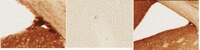
Immunohistochemistry Analysis: A 1:1,000 dilution of this antibody immunostained neuroblasts in subventricular zone (SVZ) of the lateral ventricle (LV) of 4% paraformaldehyde-fixed free-floating C57BL/6 mouse brain sections.
Usage Statement
Unless otherwise stated in our catalog or other company documentation accompanying the product(s), our products are intended for research use only and are not to be used for any other purpose, which includes but is not limited to, unauthorized commercial uses, in vitro diagnostic uses, ex vivo or in vivo therapeutic uses or any type of consumption or application to humans or animals.
Storage and Shipping Information
Storage Conditions
Stable for 1 year at -20°C from date of receipt.
Handling Recommendations: Upon receipt and prior to removing the cap, centrifuge the vial and gently mix the solution. Aliquot into microcentrifuge tubes and store at -20°C. Avoid repeated freeze/thaw cycles, which may damage IgG and affect product performance.