Wenn Sie das Fenster schließen, wird Ihre Konfiguration nicht gespeichert, es sei denn, Sie haben Ihren Artikel in die Bestellung aufgenommen oder zu Ihren Favoriten hinzugefügt.
Klicken Sie auf OK, um das MILLIPLEX® MAP-Tool zu schließen oder auf Abbrechen, um zu Ihrer Auswahl zurückzukehren.
Wählen Sie konfigurierbare Panels & Premixed-Kits - ODER - Kits für die zelluläre Signaltransduktion & MAPmates™
Konfigurieren Sie Ihre MILLIPLEX® MAP-Kits und lassen sich den Preis anzeigen.
Konfigurierbare Panels & Premixed-Kits
Unser breites Angebot enthält Multiplex-Panels, für die Sie die Analyten auswählen können, die am besten für Ihre Anwendung geeignet sind. Unter einem separaten Register können Sie das Premixed-Cytokin-Format oder ein Singleplex-Kit wählen.
Kits für die zelluläre Signaltransduktion & MAPmates™
Wählen Sie gebrauchsfertige Kits zur Erforschung gesamter Signalwege oder Prozesse. Oder konfigurieren Sie Ihre eigenen Kits mit Singleplex MAPmates™.
Die folgenden MAPmates™ sollten nicht zusammen analysiert werden: -MAPmates™, die einen unterschiedlichen Assaypuffer erfordern. -Phosphospezifische und MAPmate™ Gesamtkombinationen wie Gesamt-GSK3β und Gesamt-GSK3β (Ser 9). -PanTyr und locusspezifische MAPmates™, z.B. Phospho-EGF-Rezeptor und Phospho-STAT1 (Tyr701). -Mehr als 1 Phospho-MAPmate™ für ein einziges Target (Akt, STAT3). -GAPDH und β-Tubulin können nicht mit Kits oder MAPmates™, die panTyr enthalten, analysiert werden.
.
Bestellnummer
Bestellinformationen
St./Pkg.
Liste
Dieser Artikel wurde zu Ihren Favoriten hinzugefügt.
Wählen Sie bitte Spezies, Panelart, Kit oder Probenart
Um Ihr MILLIPLEX® MAP-Kit zu konfigurieren, wählen Sie zunächst eine Spezies, eine Panelart und/oder ein Kit.
Custom Premix Selecting "Custom Premix" option means that all of the beads you have chosen will be premixed in manufacturing before the kit is sent to you.
Catalogue Number
Ordering Description
Qty/Pack
List
Dieser Artikel wurde zu Ihren Favoriten hinzugefügt.
Spezies
Panelart
Gewähltes Kit
Menge
Bestellnummer
Bestellinformationen
St./Pkg.
Listenpreis
96-Well Plate
Menge
Bestellnummer
Bestellinformationen
St./Pkg.
Listenpreis
Weitere Reagenzien hinzufügen (MAPmates erfordern die Verwendung eines Puffer- und Detektionskits)
Menge
Bestellnummer
Bestellinformationen
St./Pkg.
Listenpreis
48-602MAG
Buffer Detection Kit for Magnetic Beads
1 Kit
Platzsparende Option Kunden, die mehrere Kits kaufen, können ihre Multiplex-Assaykomponenten in Kunststoffbeuteln anstelle von Packungen erhalten, um eine kompaktere Lagerung zu ermöglichen.
Dieser Artikel wurde zu Ihren Favoriten hinzugefügt.
Das Produkt wurde in Ihre Bestellung aufgenommen
Sie können nun ein weiteres Kit konfigurieren, ein Premixed-Kit wählen, zur Kasse gehen oder das Bestell-Tool schließen.
Anti-Parkin Antibody, clone PRK8 detects level of Parkin & has been published & validated for use in IP, WB, IC, IH.
More>>Anti-Parkin Antibody, clone PRK8 detects level of Parkin & has been published & validated for use in IP, WB, IC, IH. Less<<
Anti-Parkin Antibody, clone PRK8: SDB (Sicherheitsdatenblätter), Analysenzertifikate und Qualitätszertifikate, Dossiers, Broschüren und andere verfügbare Dokumente.
The precise function of this gene is unknown; however, the encoded protein is a component of a multiprotein E3 ubiquitin ligase complex that mediates the targeting of substrate proteins for proteasomal degradation. Mutations in this gene are known to cause Parkinson disease and autosomal recessive juvenile Parkinson disease. Alternative splicing of this gene produces multiple transcript variants encoding distinct isoforms. Additional splice variants of this gene have been described but currently lack transcript support.
FUNCTION: SwissProt: O60260 # Functions within a multiprotein E3 ubiquitin ligase complex, catalyzing the covalent attachment of ubiquitin moieties onto substrate proteins. These substrates include SYT11, CCNE1, GPR37, STUB1, a 22 kDa O-linked glycosylated isoform of SNCAIP and SEPT5. May play a more general role in the ubiquitin proteasomal pathway by participating in the removal and/or detoxification of abnormally folded or damaged protein. Loss of this ubiquitin ligase activity appears to be the mechanism underlying pathogenesis of PARK2. May protect neurons against alpha synuclein toxicity, proteasomal dysfunction, GPR37 accumulation, and kainate-induced excitotoxicity. May play a role in controlling neurotransmitter trafficking at the presynaptic terminal and in calcium-dependent exocytosis. Regulates cyclin E during neuronal apoptosis. May represent a tumor suppressor gene. SIZE: 465 amino acids; 51641 Da SUBUNIT: Forms an E3 ubiquitin ligase complex with UBE2L3 or UBE2L6. Part of a SCF-like complex, consisting of PARK2, CUL1 and FBXW7. Interacts with SNCAIP. Binds to the C2A and C2B domains of SYT11. Interacts and regulates the turnover of SEPT5. Part of a complex, including STUB1, HSP70 and GPR37. The amount of STUB1 in the complex increases during ER stress. STUB1 promotes the dissociation of HSP70 from PARK2 and GPR37, thus facilitating PARK2-mediated GPR37 ubiquitination. HSP70 transiently associates with unfolded GPR37 and inhibits the E3 activity of PARK2, whereas, STUB1 enhances the E3 activity of PARK2 through promotion of dissociation of HSP70 from PARK2-GPR37 complexes. Interacts with PSMD4 and PACRG. Interacts with LRRK2. Interacts with RANBP2. Interacts with SUMO1 but not SUMO2, which promotes nuclear localization and autoubiquitination. SUBCELLULAR LOCATION: Cytoplasm. Note=Co-localizes with STY11 in neutrites. Co-localizes with SNCAIP in brainstem Lewy bodies. Nucleus. TISSUE SPECIFICITY: Highly expressed in the brain including the substantia nigra. Expressed in heart, testis and skeletal muscle. Expression is down-regulated or absent in tumor biopsies, and absent in the brain of PARK2 patients. Overexpression protects dopamine neurons from kainate-mediated apoptosis. DOMAIN: SwissProt: O60260 The ubiquitin-like domain binds the PSMD4 subunit of 26S proteasomes. PTM: Auto-ubiquitinates in an E2-dependent manner leading to its own degradation. & S-nitrosylated. The inhibition of PARK2 ubiquitin E3 ligase activity by S-nitrosylation could contribute to the degenerative process in PD by impairing the ubiquitination of PARK2 substrates. DISEASE: SwissProt: O60260 # Defects in PARK2 are a cause of Parkinson disease (PD) [MIM:168600]. PD is a complex, multifactorial disorder that typically manifests after the age of 50 years, although early- onset cases (before 50 years) are known. PD generally arises as a sporadic condition but is occasionally inherited as a simple mendelian trait. Although sporadic and familial PD are very similar, inherited forms of the disease usually begin at earlier ages and are associated with atypical clinical features. PD is characterized by bradykinesia, resting tremor, muscular rigidity and postural instability, as well as by a clinically significant response to treatment with levodopa. The pathology of PD involves the loss of dopaminergic neurons in the substantia nigra and the presence of Lewy bodies (intraneuronal accumulations of aggregated proteins), in surviving neurons in various areas of the brain. & Defects in PARK2 are the cause of autosomal recessive early onset Parkinson disease 2 (PARK2) [MIM:600116]; also known as early-onset parkinsonism with diurnal fluctuation (EPDF) or autosomal recessive juvenile Parkinson disease (PDJ). PARK2 is symptomatically different in several aspects from idiopathic Parkinson disease, although classic symptoms such as bradykinesia, rigidity and tremor are present. Additional clinical features include early DOPA-induced dyskinesia, diurnal fluctuation of the symptoms, sleep benefit, dystonia and hyper-reflexia. PARK2 is usually characterized by onset before 40, with a mean age at onset of 23.2 years. Pathologically, PARK2 patients show loss of dopaminergic neurons in the substantia nigra, similar to that seen in Parkinson disease; however, Lewy bodies (intraneuronal accumulations of aggregated proteins) are absent. & Defects in PARK2 may be involved in the development and/or progression of ovarian cancer. SIMILARITY: Contains 2 IBR-type zinc fingers. & Contains 2 RING-type zinc fingers. & Contains 1 ubiquitin-like domain. MISCELLANEOUS: The parkin locus (PRKN), adjacent to the 6q telomere is hyper-recombinable and lies within FRA6E, the third most common fragile site in tumor tissue.
Molecular Weight
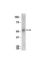
52kDa. May be observed as a doublet at 52 and 46kDa
Physicochemical Information
Dimensions
Materials Information
Toxicological Information
Safety Information according to GHS
Safety Information
Product Usage Statements
Quality Assurance
routinely evaluated by immunoblot on RIPA lysates from HEK293 cells
Usage Statement
Unless otherwise stated in our catalog or other company documentation accompanying the product(s), our products are intended for research use only and are not to be used for any other purpose, which includes but is not limited to, unauthorized commercial uses, in vitro diagnostic uses, ex vivo or in vivo therapeutic uses or any type of consumption or application to humans or animals.
Cardiomyocyte hypertrophy has been associated with diminished mitochondrial metabolism. Mitochondria are crucial organelles for the production of ATP, and their morphology and function are regulated by the dynamic processes of fusion and fission. The relationship between mitochondrial dynamics and cardiomyocyte hypertrophy is still poorly understood. Here, we show that treatment of cultured neonatal rat cardiomyocytes with the hypertrophic agonist norepinephrine promotes mitochondrial fission (characterized by a decrease in mitochondrial mean volume and an increase in the relative number of mitochondria per cell) and a decrease in mitochondrial function. We demonstrate that norepinephrine acts through α1-adrenergic receptors to increase cytoplasmic Ca(2+), activating calcineurin and promoting migration of the fission protein Drp1 (encoded by Dnml1) to mitochondria. Dominant-negative Drp1 (K38A) not only prevented mitochondrial fission, it also blocked hypertrophic growth of cardiomyocytes in response to norepinephrine. Remarkably, an antisense adenovirus against the fusion protein Mfn2 (AsMfn2) was sufficient to increase mitochondrial fission and stimulate a hypertrophic response without agonist treatment. Collectively, these results demonstrate the importance of mitochondrial dynamics in the development of cardiomyocyte hypertrophy and metabolic remodeling.
CONTEXT: The cause and pathogenesis of Parkinson's disease remain unknown; mitochondrial dysfunction, oxidative damage, environmental factors, and genetic predisposition might all be involved. Identification of the causative genes for familial Parkinson's diseases allow study of the pathogenesis of the disease at the molecular level. STARTING POINT: Katja Hedrich and colleagues studied 75 Serbian patients with early-onset Parkinson's disease for DJ-1 mutations (Neurology 2004; 62: 389-94). One patient was a compound heterozygote and another had a heterozygous exon deletion. DJ-1 mutations seem to be rare in this European population. By contrast, parkin mutations have been found in about 50% of familial cases and in 10-20% of cases without a positive family history. WHERE NEXT: The fact that parkin is a ubiquitin ligase gives special meaning to the molecular mechanism of neurodegeneration in general. In Parkinson's disease, Lewy bodies are immunoreactive for ubiquitin. Accumulation of abnormal proteins has also been seen in other neurodegenerative disorders. Disturbance of protein degradation by the ubiquitin-proteasome system might have a critical role in neurodegeneration. Although alpha-synuclein mutations are infrequent, alpha-synuclein accumulates in Lewy bodies, and alpha-synuclein fibrils impair the 26S proteasome function. UCH-L1 is also an abundant deubiquitylating enzyme, and its mutation is linked to PARK5. Furthermore, DJ-1 might interact with SUMO-1 (small ubiquitin-like modifier), which can counteract ubiquitin and stabilise proteins against degradation by the 26S proteasome. Uncovering the mechanisms of protein degradation should add importantly to understanding the neurodegenerative process in these neurodegenerative diseases.
The ubiquitin-proteasome system (UPS) is important for intracellular proteolysis, and is responsible for a diverse array of biologically important cellular processes, such as cell-cycle progression, signaling cascades and developmental programs. This system is also involved in the protein quality control, which maintains the health of the cell. Thus, the UPS provides a clue for understanding of the molecular mechanisms underlying various neurodegenerative diseases. In the last decade, we witnessed a tremendous progress in uncovering the mechanisms of Parkinson's disease (PD). Of the several genes that can cause familial PD, parkin, the causative gene of autosomal recessive juvenile parkinsonism (ARJP), is of a special interest because it encodes an ubiquitin-protein ligase, which covalently attaches ubiquitin to target proteins, designating them for destruction by the proteasome. This review summarizes recent studies on the UPS pathway with a special reference to parkin, focusing on how parkin is linked to the pathogenesis of ARJP.
Parkinson's disease (PD) is the most common neurodegenerative movement disorder. The major motor disabilities of PD are associated with the extensive loss of dopaminergic neurons in the substantia nigra pars compacta. The physiological changes and biochemical pathways involved in the selective demise of these neurons are still unclear. Recent studies have demonstrated that alterations or reductions in ubiquitin-mediated proteasome function can be causal of at least some forms of parkinsonism, and multiple lines of evidence suggest that this mechanism of protein degradation may play an important role in the etiology of PD.
Kindreds with Mendelian inheritance of Parkinson disease (PD) have been known since a long time ago. Nine loci have been mapped in familial PD by linkage study and four causative genes have been cloned. This paper discusses Park 1 and Park 2, the identification of which has brought about many advances in the studies on pathomechanism of PD. Investigations of these genes in familial PD have expanded their clinical and pathological phenotypes. However, to clarify the effect of mutations on these phenotypes, additional post-mortem neuropathological studies are required.
Parkinson's disease (PD) is a complex disorder with many different causes, yet they may intersect in common pathways, raising the possibility that neuroprotective agents may have broad applicability in the treatment of PD. Current evidence suggests that mitochondrial complex I inhibition may be the central cause of sporadic PD and that derangements in complex I cause alpha-synuclein aggregation, which contributes to the demise of dopamine neurons. Accumulation and aggregation of alpha-synuclein may further contribute to the death of dopamine neurons through impairments in protein handling and detoxification. Dysfunction of parkin (a ubiquitin E3 ligase) and DJ-1 could contribute to these deficits. Strategies aimed at restoring complex I activity, reducing oxidative stress and alpha-synuclein aggregation, and enhancing protein degradation may hold particular promise as powerful neuroprotective agents in the treatment of PD.