Wenn Sie das Fenster schließen, wird Ihre Konfiguration nicht gespeichert, es sei denn, Sie haben Ihren Artikel in die Bestellung aufgenommen oder zu Ihren Favoriten hinzugefügt.
Klicken Sie auf OK, um das MILLIPLEX® MAP-Tool zu schließen oder auf Abbrechen, um zu Ihrer Auswahl zurückzukehren.
Wählen Sie konfigurierbare Panels & Premixed-Kits - ODER - Kits für die zelluläre Signaltransduktion & MAPmates™
Konfigurieren Sie Ihre MILLIPLEX® MAP-Kits und lassen sich den Preis anzeigen.
Konfigurierbare Panels & Premixed-Kits
Unser breites Angebot enthält Multiplex-Panels, für die Sie die Analyten auswählen können, die am besten für Ihre Anwendung geeignet sind. Unter einem separaten Register können Sie das Premixed-Cytokin-Format oder ein Singleplex-Kit wählen.
Kits für die zelluläre Signaltransduktion & MAPmates™
Wählen Sie gebrauchsfertige Kits zur Erforschung gesamter Signalwege oder Prozesse. Oder konfigurieren Sie Ihre eigenen Kits mit Singleplex MAPmates™.
Die folgenden MAPmates™ sollten nicht zusammen analysiert werden: -MAPmates™, die einen unterschiedlichen Assaypuffer erfordern. -Phosphospezifische und MAPmate™ Gesamtkombinationen wie Gesamt-GSK3β und Gesamt-GSK3β (Ser 9). -PanTyr und locusspezifische MAPmates™, z.B. Phospho-EGF-Rezeptor und Phospho-STAT1 (Tyr701). -Mehr als 1 Phospho-MAPmate™ für ein einziges Target (Akt, STAT3). -GAPDH und β-Tubulin können nicht mit Kits oder MAPmates™, die panTyr enthalten, analysiert werden.
.
Bestellnummer
Bestellinformationen
St./Pkg.
Liste
Dieser Artikel wurde zu Ihren Favoriten hinzugefügt.
Wählen Sie bitte Spezies, Panelart, Kit oder Probenart
Um Ihr MILLIPLEX® MAP-Kit zu konfigurieren, wählen Sie zunächst eine Spezies, eine Panelart und/oder ein Kit.
Custom Premix Selecting "Custom Premix" option means that all of the beads you have chosen will be premixed in manufacturing before the kit is sent to you.
Catalogue Number
Ordering Description
Qty/Pack
List
Dieser Artikel wurde zu Ihren Favoriten hinzugefügt.
Spezies
Panelart
Gewähltes Kit
Menge
Bestellnummer
Bestellinformationen
St./Pkg.
Listenpreis
96-Well Plate
Menge
Bestellnummer
Bestellinformationen
St./Pkg.
Listenpreis
Weitere Reagenzien hinzufügen (MAPmates erfordern die Verwendung eines Puffer- und Detektionskits)
Menge
Bestellnummer
Bestellinformationen
St./Pkg.
Listenpreis
48-602MAG
Buffer Detection Kit for Magnetic Beads
1 Kit
Platzsparende Option Kunden, die mehrere Kits kaufen, können ihre Multiplex-Assaykomponenten in Kunststoffbeuteln anstelle von Packungen erhalten, um eine kompaktere Lagerung zu ermöglichen.
Dieser Artikel wurde zu Ihren Favoriten hinzugefügt.
Das Produkt wurde in Ihre Bestellung aufgenommen
Sie können nun ein weiteres Kit konfigurieren, ein Premixed-Kit wählen, zur Kasse gehen oder das Bestell-Tool schließen.
Anti-SREBP-2, clone 22D5, Cat. No. MAB1988, a rabbit monoclonal antibody detects murine SREBP-2 by Western Blotting.
More>>Anti-SREBP-2, clone 22D5, Cat. No. MAB1988, a rabbit monoclonal antibody detects murine SREBP-2 by Western Blotting. Less<<
Anti-SREBP-2 Antibody, clone 22D5: SDB (Sicherheitsdatenblätter), Analysenzertifikate und Qualitätszertifikate, Dossiers, Broschüren und andere verfügbare Dokumente.
Sterol regulatory element-binding protein 2 (UniProt: Q3U1N2; also known as SREBP-2, Sterol regulatory element-binding transcription factor 2) is encoded by the Srebf2 (also known as Srebp2) gene (Gene ID: 20788) in murine species. SREBPs are a family of transcription factors that regulate lipid homeostasis by controlling the expression of a range of enzymes that are required for endogenous cholesterol, fatty acid, triacylglycerol, and phospholipid synthesis. The three SREBP isoforms known as, SREBP-1a, SREBP-1c, and SREBP-2, have different roles in lipid synthesis. SREBP-1 and SREBP-2 proteins share 47% of homology. SREBP-2 is mainly involved in cholesterol synthesis and SREBP-1c is mainly involved in fatty acid synthesis and insulin induced glucose metabolism and SREBP-1a isoform is involved in both of these pathways. SREBPs are synthetized as inactive precursor proteins that are bound to the endoplasmic reticulum membranes and upon activation, the precursor is cleaved off in a two-step process to release the N-terminal active domain in the nucleus. SREBP precursors are organized into three domains - an N-terminal domain that contains the transactivation domain, a region rich in serine and proline, and the bHLH-LZ region for DNA binding and dimerization. Sterols are shown to inhibit the cleavage of the precursor protein and the mature nuclear form is rapidly catabolized, thereby reducing transcription. It regulates transcription of the LDL receptor gene as well as the cholesterol and to a lesser degree the fatty acid synthesis pathway. It binds the sterol regulatory element 1 (SRE-1) (5'-ATCACCCCAC-3') found in the flanking region of the LDRL and HMG-CoA synthase genes. The hepatic overexpression of SREBP-2 isoform in mice causes a preferential induction of genes involved in cholesterol biosynthesis. (Ref.: Eberle, D et al. (2004) Biochimie 86(11); 839-48).
References
Product Information
Format
Unpurified
Presentation
Rabbit monoclonal antibody in cell culture supernatant without azide.
Anti-SREBP-2, clone 22D5, Cat. No. MAB1988, a rabbit monoclonal antibody detects murine SREBP-2 by Western Blotting.
Key Applications
Western Blotting
Application Notes
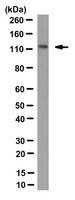
Western Blotting Analysis: A 1:1,000 dilution from a representative lot detected SREBP-2 in 10 µg of HEK293 and HeLa cell lysates.
Western Blotting Analysis: A representative lot detected SREBP-2 in Western Blotting applications (McFarlane, M.R., et. al. (2015). J Lipid Res. 56(8):1560-71).
Biological Information
Immunogen
His-tagged recombinant fragment corresponding to 219 amino acids from the N-terminal region of murine Sterol regulatory element-binding protein 2.
Clone
22D5
Concentration
Please refer to lot specific datasheet.
Host
Rabbit
Specificity
Clone 22D5 specifically detected Sterol regulatory element-binding protein 2. It targets an epitope within the 219 amino acids from the N-terminal region.
~122 kDa observed; 122.91 kDa calculated. Uncharacterized bands may be observed in some lysate(s).
Physicochemical Information
Dimensions
Materials Information
Toxicological Information
Safety Information according to GHS
Safety Information
Product Usage Statements
Quality Assurance
Evaluated by Western Blotting in HepG2 cell lysate.
Western Blotting Analysis: A 1:1,000 dilution of this antibody detected SREBP-2 in 10 µg of HepG2 cell lysate.
Usage Statement
Unless otherwise stated in our catalog or other company documentation accompanying the product(s), our products are intended for research use only and are not to be used for any other purpose, which includes but is not limited to, unauthorized commercial uses, in vitro diagnostic uses, ex vivo or in vivo therapeutic uses or any type of consumption or application to humans or animals.
Storage and Shipping Information
Storage Conditions
Stable for 1 year at -20°C from date of receipt.
Handling Recommendations: Upon receipt and prior to removing the cap, centrifuge the vial and gently mix the solution. Aliquot into microcentrifuge tubes and store at -20°C. Avoid repeated freeze/thaw cycles, which may damage IgG and affect product performance.