Wenn Sie das Fenster schließen, wird Ihre Konfiguration nicht gespeichert, es sei denn, Sie haben Ihren Artikel in die Bestellung aufgenommen oder zu Ihren Favoriten hinzugefügt.
Klicken Sie auf OK, um das MILLIPLEX® MAP-Tool zu schließen oder auf Abbrechen, um zu Ihrer Auswahl zurückzukehren.
Wählen Sie konfigurierbare Panels & Premixed-Kits - ODER - Kits für die zelluläre Signaltransduktion & MAPmates™
Konfigurieren Sie Ihre MILLIPLEX® MAP-Kits und lassen sich den Preis anzeigen.
Konfigurierbare Panels & Premixed-Kits
Unser breites Angebot enthält Multiplex-Panels, für die Sie die Analyten auswählen können, die am besten für Ihre Anwendung geeignet sind. Unter einem separaten Register können Sie das Premixed-Cytokin-Format oder ein Singleplex-Kit wählen.
Kits für die zelluläre Signaltransduktion & MAPmates™
Wählen Sie gebrauchsfertige Kits zur Erforschung gesamter Signalwege oder Prozesse. Oder konfigurieren Sie Ihre eigenen Kits mit Singleplex MAPmates™.
Die folgenden MAPmates™ sollten nicht zusammen analysiert werden: -MAPmates™, die einen unterschiedlichen Assaypuffer erfordern. -Phosphospezifische und MAPmate™ Gesamtkombinationen wie Gesamt-GSK3β und Gesamt-GSK3β (Ser 9). -PanTyr und locusspezifische MAPmates™, z.B. Phospho-EGF-Rezeptor und Phospho-STAT1 (Tyr701). -Mehr als 1 Phospho-MAPmate™ für ein einziges Target (Akt, STAT3). -GAPDH und β-Tubulin können nicht mit Kits oder MAPmates™, die panTyr enthalten, analysiert werden.
.
Bestellnummer
Bestellinformationen
St./Pkg.
Liste
Dieser Artikel wurde zu Ihren Favoriten hinzugefügt.
Wählen Sie bitte Spezies, Panelart, Kit oder Probenart
Um Ihr MILLIPLEX® MAP-Kit zu konfigurieren, wählen Sie zunächst eine Spezies, eine Panelart und/oder ein Kit.
Custom Premix Selecting "Custom Premix" option means that all of the beads you have chosen will be premixed in manufacturing before the kit is sent to you.
Catalogue Number
Ordering Description
Qty/Pack
List
Dieser Artikel wurde zu Ihren Favoriten hinzugefügt.
Spezies
Panelart
Gewähltes Kit
Menge
Bestellnummer
Bestellinformationen
St./Pkg.
Listenpreis
96-Well Plate
Menge
Bestellnummer
Bestellinformationen
St./Pkg.
Listenpreis
Weitere Reagenzien hinzufügen (MAPmates erfordern die Verwendung eines Puffer- und Detektionskits)
Menge
Bestellnummer
Bestellinformationen
St./Pkg.
Listenpreis
48-602MAG
Buffer Detection Kit for Magnetic Beads
1 Kit
Platzsparende Option Kunden, die mehrere Kits kaufen, können ihre Multiplex-Assaykomponenten in Kunststoffbeuteln anstelle von Packungen erhalten, um eine kompaktere Lagerung zu ermöglichen.
Dieser Artikel wurde zu Ihren Favoriten hinzugefügt.
Das Produkt wurde in Ihre Bestellung aufgenommen
Sie können nun ein weiteres Kit konfigurieren, ein Premixed-Kit wählen, zur Kasse gehen oder das Bestell-Tool schließen.
ABN2180-100UL
Sigma-AldrichAnti-Synaptotagmin-1
Anti-Synaptotagmin-1, Cat. No. ABN2180, is a rabbit polyclonal antibody that detects Synaptotagmin-1 and is tested for use in Activity Assay, Immunocytochemistry, Immunofluorescence, and Western Blotting.
More>>Anti-Synaptotagmin-1, Cat. No. ABN2180, is a rabbit polyclonal antibody that detects Synaptotagmin-1 and is tested for use in Activity Assay, Immunocytochemistry, Immunofluorescence, and Western Blotting. Less<<
Synaptotagmin-1 (UniProt: P21707; also known as Synaptotagmin I, SytI, p65) is encoded by the Syt1 gene (Gene ID:25716) in rat. Synaptotagmin-1 is a single-pass homotetrameric membrane protein that serves as a calcium sensor and participates in triggering neurotransmitter release at the synapse. It is one of the major synaptic vesicle proteins with a vesicular domain (aa 1-57), a transmembrane domain (aa 58-79), and a cytoplasmic domain (aa 80-421). The cytoplasmic domain contains two calcium-binding motifs (C2A; aa 141-260 and C2B; aa 272-405) that bind a total of five calcium ions three by C2A and two by C2B. C2A mediates calcium-dependent phospholipid binding and C2B mediates the interaction with synaptic vesicle protein 2A (SV2A) and stoning 2. Synaptotagmin-1 plays a regulatory role in membrane interactions during trafficking of synaptic vesicles at the active zone of the synapse. It binds acidic phospholipids with a specificity that requires the presence of both an acidic head group and a diacyl backbone. A calcium-dependent interaction between synaptotagmin and putative receptors for activated protein kinase C has also been reported. (Ref.: Malgaroli, A., et al. (1995). Science 268 (5217), 1624-1628).
References
Product Information
Format
Purified
Presentation
Purified rabbit polyclonal antibody in buffer containing 0.1 M Tris-Glycine (pH 7.4), 150 mM NaCl with 0.05% sodium azide.
Anti-Synaptotagmin-1, Cat. No. ABN2180, is a rabbit polyclonal antibody that detects Synaptotagmin-1 and is tested for use in Activity Assay, Immunocytochemistry, Immunofluorescence, and Western Blotting.
Key Applications
Activity Assay
Immunocytochemistry
Immunofluorescence
Western Blotting
Application Notes
Immunocytochemistry Analysis: A representative lot detected Synaptotagmin-1 in Immunocytochemistry applications (Malgaroli, A., et. al. (1995). Science. 268(5217):1624-8).
Western Blotting Analysis: A representative lot detected Synaptotagmin-1 in Western Blotting applications (Malgaroli, A., et. al. (1995). Science. 268(5217):1624-8).
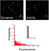
Immunofluorescence Analysis: A 1:200 dilution from a representative lot detected Synaptotagmin-1 in Cultured Rat Hippocampal cells (Courtesy of Dr. Antonio Malgaroli @ Universita' Vita-Salute San Raffaele, Milano, Italy).
Activity Assay: A representative lot was used to detect changes in synaptic activity. (Malgaroli, A., et. al. (1995). Science. 268(5217):1624-8).
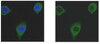
Immunofluorescence Analysis: A representative lot detected Synaptotagmin-1 in Immunofluorescence applications (Ferro, M., et. al. (2017). Nat Commun. 8(1):1229).
Western Blotting Analysis: A 1:500 dilution from a representative lot detected Synaptotagmin-1 in Rat Brain Cortex (Courtesy of Dr. Antonio Malgaroli @ Universita' Vita-Salute San Raffaele, Milano, Italy).
Note: Actual optimal working dilutions must be determined by end user as specimens, and experimental conditions may vary with the end user
Biological Information
Immunogen
BSA-conjugated linear peptide corresponding to the first 23 amino acids from the N-terminal vesicular domain of rat Synaptotagmin-1.
Epitope
N-terminus
Concentration
0.5 mg/mL. Please refer to guidance on suggested starting dilutions and/or titers per application and sample type.
Host
Rabbit
Specificity
This rabbit polyclonal antibody detects rat Synaptotagmin-1. It targets an epitope within the N-terminal, vesicular domain.
~58 kDa observed; 47.40 kDa calculated. Uncharacterized bands may be observed in some lysate(s).
Physicochemical Information
Dimensions
Materials Information
Toxicological Information
Safety Information according to GHS
Safety Information
Product Usage Statements
Quality Assurance
Evaluated by Western Blotting in Rat Brain Synaptosomes.
Western Blotting Analysis: A 1:1,000 dilution of this antibody detected Synaptotagmin-1 in Rat Brain Synaptosomes.
Usage Statement
Unless otherwise stated in our catalog or other company documentation accompanying the product(s), our products are intended for research use only and are not to be used for any other purpose, which includes but is not limited to, unauthorized commercial uses, in vitro diagnostic uses, ex vivo or in vivo therapeutic uses or any type of consumption or application to humans or animals.
Storage and Shipping Information
Storage Conditions
Stable for 1 year at +2°C to +8°C from date of receipt.
Ideally, elucidating the role of specific brain circuits in animal behavior would require the ability to measure activity at all involved synapses, possibly with unrestricted field of view, thus even at those boutons deeply located into the brain. Here, we introduce and validate an efficient scheme reporting synaptic vesicle cycling in vivo. This is based on SynaptoZip, a genetically encoded molecule deploying in the vesicular lumen a bait moiety designed to capture upon exocytosis a labeled alien peptide, Synbond. The resulting signal is cumulative and stores the number of cycling events occurring at individual synapses. Since this functional signal is enduring and measurable both online and ex post, SynaptoZip provides a unique method for the analysis of the history of synaptic activity in regions several millimeters below the brain surface. We show its broad applicability by reporting stimulus-evoked and spontaneous circuit activity in wide cortical fields, in anesthetized and freely moving animals.
Presynaptic component of long-term potentiation visualized at individual hippocampal synapses A Malgaroli 1 , A E Ting, B Wendland, A Bergamaschi, A Villa, R W Tsien, R H Scheller Science
268(5217)
1624-8
1994
Long-term potentiation has previously been studied with electrophysiological techniques that do not readily separate presynaptic and postsynaptic contributions. Changes in exocytotic-endocytotic cycling have now been monitored at synapses between cultured rat hippocampal neurons by measuring the differential uptake of antibodies that recognize the intraluminal domain of the synaptic vesicle protein synaptotagmin. Vesicular cycling increased markedly during glutamate-induced long-term potentiation. The degree of potentiation was heterogeneous, appearing greater at synapses at which the initial extent of vesicular turnover was low. Thus, changes in presynaptic activity were visualized directly and the spatial distribution of potentiation could be determined at the level of single synaptic boutons.