Wenn Sie das Fenster schließen, wird Ihre Konfiguration nicht gespeichert, es sei denn, Sie haben Ihren Artikel in die Bestellung aufgenommen oder zu Ihren Favoriten hinzugefügt.
Klicken Sie auf OK, um das MILLIPLEX® MAP-Tool zu schließen oder auf Abbrechen, um zu Ihrer Auswahl zurückzukehren.
Wählen Sie konfigurierbare Panels & Premixed-Kits - ODER - Kits für die zelluläre Signaltransduktion & MAPmates™
Konfigurieren Sie Ihre MILLIPLEX® MAP-Kits und lassen sich den Preis anzeigen.
Konfigurierbare Panels & Premixed-Kits
Unser breites Angebot enthält Multiplex-Panels, für die Sie die Analyten auswählen können, die am besten für Ihre Anwendung geeignet sind. Unter einem separaten Register können Sie das Premixed-Cytokin-Format oder ein Singleplex-Kit wählen.
Kits für die zelluläre Signaltransduktion & MAPmates™
Wählen Sie gebrauchsfertige Kits zur Erforschung gesamter Signalwege oder Prozesse. Oder konfigurieren Sie Ihre eigenen Kits mit Singleplex MAPmates™.
Die folgenden MAPmates™ sollten nicht zusammen analysiert werden: -MAPmates™, die einen unterschiedlichen Assaypuffer erfordern. -Phosphospezifische und MAPmate™ Gesamtkombinationen wie Gesamt-GSK3β und Gesamt-GSK3β (Ser 9). -PanTyr und locusspezifische MAPmates™, z.B. Phospho-EGF-Rezeptor und Phospho-STAT1 (Tyr701). -Mehr als 1 Phospho-MAPmate™ für ein einziges Target (Akt, STAT3). -GAPDH und β-Tubulin können nicht mit Kits oder MAPmates™, die panTyr enthalten, analysiert werden.
.
Bestellnummer
Bestellinformationen
St./Pkg.
Liste
Dieser Artikel wurde zu Ihren Favoriten hinzugefügt.
Wählen Sie bitte Spezies, Panelart, Kit oder Probenart
Um Ihr MILLIPLEX® MAP-Kit zu konfigurieren, wählen Sie zunächst eine Spezies, eine Panelart und/oder ein Kit.
Custom Premix Selecting "Custom Premix" option means that all of the beads you have chosen will be premixed in manufacturing before the kit is sent to you.
Catalogue Number
Ordering Description
Qty/Pack
List
Dieser Artikel wurde zu Ihren Favoriten hinzugefügt.
Spezies
Panelart
Gewähltes Kit
Menge
Bestellnummer
Bestellinformationen
St./Pkg.
Listenpreis
96-Well Plate
Menge
Bestellnummer
Bestellinformationen
St./Pkg.
Listenpreis
Weitere Reagenzien hinzufügen (MAPmates erfordern die Verwendung eines Puffer- und Detektionskits)
Menge
Bestellnummer
Bestellinformationen
St./Pkg.
Listenpreis
48-602MAG
Buffer Detection Kit for Magnetic Beads
1 Kit
Platzsparende Option Kunden, die mehrere Kits kaufen, können ihre Multiplex-Assaykomponenten in Kunststoffbeuteln anstelle von Packungen erhalten, um eine kompaktere Lagerung zu ermöglichen.
Dieser Artikel wurde zu Ihren Favoriten hinzugefügt.
Das Produkt wurde in Ihre Bestellung aufgenommen
Sie können nun ein weiteres Kit konfigurieren, ein Premixed-Kit wählen, zur Kasse gehen oder das Bestell-Tool schließen.
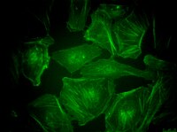
SCT215
Sigma-AldrichBioTracker™ 497 Green Actin Live Cell Probe
Live cell imaging cytoskeleton probe used to detect actin using fluorescence microscopy or flow cytometry.
More>>Live cell imaging cytoskeleton probe used to detect actin using fluorescence microscopy or flow cytometry. Less<<
Empfohlene Produkte
Übersicht
Replacement Information
Description
Catalogue Number
SCT215
Description
BioTracker™ 497 Green Actin Live Cell Probe
Overview
The BioTracker™ 497 Green Actin Live Cell Probe may be used in both living cells and fixed-cell applications. Because SCT215 is an organic fluorescent small molecule with ready membrane permeability, it enables visualization of intracellular actin filamentsby simple addition of the probe to the cell culture medium or extracellular fluid without a rinsing step.
This probe is thought to bind to F-actin through the common binding site for phalloidin and jasplakinolide.
The BioTracker™ 497 Green Actin Live Cell Probe is a fluorescent probe that specifically binds to actin filaments (F-actin) and can visualize F-actin with green fluorescence.
Live cell imaging cytoskeleton probe used to detect actin using fluorescence microscopy or flow cytometry.
Key Applications
Cell Based Assays
Flow Cytometry
Fluorescence
Application Notes
SCT215 fluorescence can be observed with a blue excitation and green fluorescence filter set for GFP/FITC. For live-cell imaging, use lower concentrations of probe, weaken the excitation intensity and use high-sensitivity cameras to reduce exposure time. Consider the use of antifade reagent for time-lapse or continuous imaging. Stock solution is prepared by adding 30 uL DMSO to one vial,, which results in a pale yellow liquid. Diluting stock solution in culture medium to 50-100 nM before adding to cells is recommended, although optimal incubation concentraiton may be determined according to user application. Use the lowest possible concentration to limit cytotoxicity.
Biological Information
Physicochemical Information
Dimensions
Materials Information
Toxicological Information
Safety Information according to GHS
Safety Information
Product Usage Statements
Usage Statement
Unless otherwise stated in our catalog or other company documentation accompanying the product(s), our products are intended for research use only and are not to be used for any other purpose, which includes but is not limited to, unauthorized commercial uses, in vitro diagnostic uses, ex vivo or in vivo therapeutic uses or any type of consumption or application to humans or animals.
Storage and Shipping Information
Storage Conditions
Store BioTracker™ 497 Green Actin Live Cell Probe at -20°C, desiccated and protected from light. Centrifuge vial briefly to collect contents at bottom of vial before opening. After dissolving in DMSO, store aliquots at -20 °C.