Si cierra, no se guardará su personalización salvo que haya añadido el artículo a su carrito de la compra o a favoritos.
Pulse OK para cerrar la herramienta MILLIPLEX® MAP o Cancelar para volver a su selección.
Elija paneles personalizables y kits premezclos - O - MAPmates™ de señalización celular
Diseñe y calcule el precio de sus kits MILLIPLEX® MAP.
Paneles personalizados y kits premezclados
Nuestra amplia cartera de productos consta de paneles multiplex que le permiten elegir, dentro del panel, los analitos que mejor se ajustan a sus requisitos. En una pestaña distinta puede elegir el formato de citocina premezclada o un kit single plex.
Kits de señalización celular y MAPmates™
Elija los kits preparados para poder explorar las vías o los procesos enteros. O diseñe sus propios kits eligiendo single plex MAPmates™ según las directrices proporcionadas.
No deben combinarse los siguientes MAPmates™: -MAPmates™ que requieren un tampón de ensayo diferente. -Pares MAPmate™ fosfoespecíficos y totales, por ejemplo, GSK3β y GSK3β (Ser 9). -MAPmates™ con panTyr y específicos de sitio; por ejemplo, receptor del fosfo-EGF y fosfo-STAT1 (Tyr701). -Más de 1 fosfo-MAPmate™ para una sola diana (Akt, STAT3). -La GAPDH y la β-tubulina no pueden combinarse con kits o MAPmates™ que contengan panTyr.
.
Número de referencia
Descripción para pedidos
Cant./Env.
Lista
Este artículo se ha añadido a favoritos.
Seleccione una especie, un tipo de panel, un kit o un tipo de muestra
Para empezar a diseñar su kit MILLIPLEX® MAP, seleccione una especie, un tipo de panel o un kit de interés.
Custom Premix Selecting "Custom Premix" option means that all of the beads you have chosen will be premixed in manufacturing before the kit is sent to you.
Catalogue Number
Ordering Description
Qty/Pack
List
Este artículo se ha añadido a favoritos.
Especie
Tipo de panel
Kit seleccionado
Cant.
Número de referencia
Descripción para pedidos
Cant./Env.
Precio de catálogo
96-Well Plate
Cant.
Número de referencia
Descripción para pedidos
Cant./Env.
Precio de catálogo
Añadir más reactivos (Se necesita tampón y un kit de detección para usar con MAPmates)
Cant.
Número de referencia
Descripción para pedidos
Cant./Env.
Precio de catálogo
48-602MAG
Buffer Detection Kit for Magnetic Beads
1 Kit
Opción para ahorrar espacio Los clientes que adquieran múltiples kits pueden optar por ahorrar espacio de almacenamiento retirando el embalaje del kit y recibiendo los componentes de sus ensayos multiplex en bolsas de plástico para un almacenamiento más compacto.
Este artículo se ha añadido a favoritos.
El producto se ha añadido a su carrito
Ahora puede personalizar otro kit, elegir un kit premezclado, tramitarlo o cerrar la herramienta de pedidos.
GR15L
Sigma-AldrichAnti-EGFR (Ab-5) Mouse mAb (EGFR.1)
Anti-EGFR (Ab-5), mouse monoclonal, clone EGFR.1, recognizes the ~170 kDa EGF receptor protein in A431 cells. It is validated for use in FC, IF, IP, and neutralization studies.
More>>Anti-EGFR (Ab-5), mouse monoclonal, clone EGFR.1, recognizes the ~170 kDa EGF receptor protein in A431 cells. It is validated for use in FC, IF, IP, and neutralization studies. Less<<
Anti-EGFR (Ab-5) Mouse mAb (EGFR.1): Ficha de datos de seguridad (MSDS o SDS), certificado de análisis y de calidad (CoA y CoQ), expedientes, folletos y otros documentos disponibles.
Stains A431, HeLa-5 cells and other human cells but not to rodent cells. It inhibits EGF binding to its receptor and is an antagonist of in vivo EGF-stimulated tyrosine kinase activity. This antibody does not cross-react to c-neu/erb B-2. Antibody should be titrated for optimal results in individual systems.
Biological Information
Immunogen
partially purified, human EGFR from A431 cells
Immunogen
Human
Clone
EGFR.1
Host
Mouse
Isotype
IgG2b
Species Reactivity
Human
Antibody Type
Monoclonal Antibody
Physicochemical Information
Dimensions
Materials Information
Toxicological Information
Safety Information according to GHS
Safety Information
Product Usage Statements
Storage and Shipping Information
Ship Code
Ambient Temperature Only
Toxicity
Standard Handling
Storage
+2°C to +8°C
Do not freeze
Ok to freeze
Special Instructions
We recommend resuspending the lyophilized antibody with sterile PBS, pH 7.4, or sterile 20 mM Tris-saline (20 mM Tris containing 0.15 M NaCl), pH 7.4, to yield a final concentration of 100 µg/ml; product will be more stable if 0.1% sodium azide is included (do not add azide if antibody is to be used with viable cells). Lyophilized antibodies should be resuspended at 4°C with occasional gentle mixing for at least 2 h.
Packaging Information
Transport Information
Supplemental Information
Specifications
Global Trade Item Number
Número de referencia
GTIN
GR15L-100UG
04055977228250
Documentation
Anti-EGFR (Ab-5) Mouse mAb (EGFR.1) Ficha datos de seguridad (MSDS)
Anti-EGFR (Ab-5) Mouse mAb (EGFR.1) Certificados de análisis
Cargo
Número de lote
GR15L
Referencias bibliográficas
Visión general referencias
Hunter T., 1984. Nature311, 414. Reynolds, F.H., Jr., et al. 1981. Nature292, 259. Ushiro, H. and Cohen, S. 1980. J. Biol. Chem.255, 8363.
Ficha técnica
Note that this data sheet is not lot-specific and is representative of the current specifications for this product. Please consult the vial label and the certificate of analysis for information on specific lots. Also note that shipping conditions may differ from storage conditions.
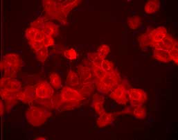
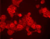
Detection of human EGFR by immunocytochemistry. Sample: A431 cells fixed with methanol. Primary Antibody: Anti-Epidermal Growth Factor Receptor (Ab-5) Mouse mAb (Cat. No. GR15L) (1 µg/ml). Detection: fluorescence.
Description
Purified mouse monoclonal antibody generated by immunizing mice with the specified immunogen and fusing splenocytes with NS-1 mouse myeloma cells. Recognizes the ~170 kDa EGF receptor protein.
Background
Epidermal growth factor (EGF) initiates its effects on cell growth through interaction with a 170,000 dalton cell surface glycoprotein receptor. Binding of EGF or transforming growth factor alpha to the receptor activates a tyrosine-specific protein kinase intrinsic to the EGF-receptor protein. The phosphotyrosine content of target cells increases rapidly and the EGF-receptor is phosphorylated on tyrosine residues.
Host
Mouse
Immunogen species
Human
Immunogen
partially purified, human EGFR from A431 cells
Clone
EGFR.1
Isotype
IgG2b
Species
human, not mouse, not rat
Positive control
A-431 cells
Negative control
HL-60 cells
Form
Lyophilized
Formulation
Lyophilized from a volatile buffer, 100 µg BSA.
Preservative
None
Comments
Stains A431, HeLa-5 cells and other human cells but not to rodent cells. It inhibits EGF binding to its receptor and is an antagonist of in vivo EGF-stimulated tyrosine kinase activity. This antibody does not cross-react to c-neu/erb B-2. Antibody should be titrated for optimal results in individual systems.
Storage
+2°C to +8°C
Do Not Freeze
Ok to freeze
Special Instructions
We recommend resuspending the lyophilized antibody with sterile PBS, pH 7.4, or sterile 20 mM Tris-saline (20 mM Tris containing 0.15 M NaCl), pH 7.4, to yield a final concentration of 100 µg/ml; product will be more stable if 0.1% sodium azide is included (do not add azide if antibody is to be used with viable cells). Lyophilized antibodies should be resuspended at 4°C with occasional gentle mixing for at least 2 h.
Toxicity
Standard Handling
References
Hunter T., 1984. Nature311, 414. Reynolds, F.H., Jr., et al. 1981. Nature292, 259. Ushiro, H. and Cohen, S. 1980. J. Biol. Chem.255, 8363.
Application references
Neutralization Studies
Gill, G., et al. 1984. J. Biol. Chem.259, 7755.
Kawamoto, T., et al. 1984. J. Biol. Chem.259, 7761.
Kawamoto, T., et al. 1983. Proc. Natl. Acad. Sci. USA80, 1337.
Sato, J.D., et al. 1983. Molec. Biol. Med.1, 511.