


Millicell® μ-Angiogenesis Activation and Inhibition Assay Kits
Recursos relacionados

Productos recomendados
-
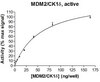
23-032M Sigma-Aldrich MDM2/CK1δ Protein, active, 250 µg -

1003990500 Supelco N,N-Dimetilacetamida -
8417470001 Sigma-Aldrich 2-Nitrofenil-ß-D-galactopiranosido -
ABE1451 Sigma-Aldrich Anti-phospho BRD4 (Ser492) -
DCSF-MH Millipore Surface finish protocol for mixing head -

ABC947-25UG Sigma-Aldrich Anti-Bif-1 -

KGW9A3TTT1 Millipore Opticap XLT30 Polysep II de 2,0/1,2 µm, TC 1 1/2 pulg. -
06-1025 Sigma-Aldrich Anti-NET39 Antibody -

EFHAW250B Millipore Unidad de filtración EZ-Fit, membrana blanca, tamaño de poro de 0,45 µm, embudo de 250 ml, envasado a granel
Descripción
Especificaciones
Información para pedidos
Documentation
Productos y aplicaciones relacionados
Productos relacionados por: Brand Facete
| Millicell® |
Categorías
| Life Science Research > Cell Analysis > Cell-based Assays > Angiogenesis & Endothelial Transmigration Assays |
Studying how compounds affect angiogenesis, either to promote or inhibit new capillary tube formation can lead to therapies affecting wound healing, tissue regeneration, cardiovascular disease, stroke, tumor progression, and more. The Millicell μ-Angiogenesis activation and inhibition kits provide a powerful, quantitative platform for real-time monitoring of changes in tubule formation with unprecedented optical resolution.
Benefits
- Continuous, live monitoring of cells as tubules form
- Even, flat slide surface prevents any liquid meniscus from forming, eliminating out-of-focus areas
- Full well visualization at low magnification
- Optimized assay conditions in a miniaturized format
- Compatible with multi-channel pipettes and fixation reagents
- Less reagents and cells needed, reducing costs and waste by up to 80% • All-in-one slide used to grow, assay, and stain cells
Supporting Data
μ-Angiogenesis Inhibition Kit
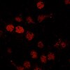
Increasing concentrations of sulforaphane resulted in both mean tube length and mean number of branch points as shown below in bright field and calcein-AM micrographs of HUVEC cells.
)
Increasing concentrations of sulforaphane resulted in both mean tube length and mean number of branch points as shown below in bright field and calcein-AM micrographs of HUVEC cells.
μ-Angiogenesis Activation Kit
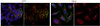
(Left to right) Bright field, calcein-AM, DAPI, and calcein-DAPI merge micrographs show robust tubule formation and lumen structure in the presence of phorbol myristate acetate (PMA, a pro-angiogenic agent). Images shown are of HUVEC cells.
)
With no stimulant
)
With 50ng/mL PMA and 1x ITS
)
Lumen structure
Applications
Angiogenesis Assays